A Map of Brain Aging express
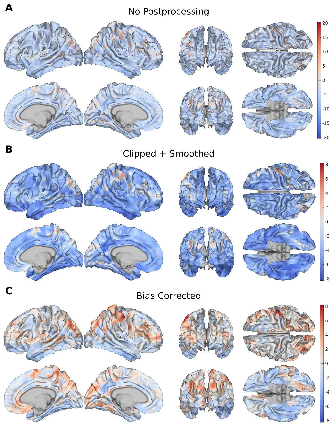
The cerebral cortex is like a crumpled sheet of paper: it has thickness, curves, and folds of varying depth. With age, this sheet changes unevenly — thinning in some places, losing the sharpness of its folds in others. Previously, doctors assessed brain age as a whole, as if looking at the sheet from a distance. Now, a neural network examines every point on the surface with a step of 1.37 mm, like a magnifying glass that reveals the aging of individual regions.
The model was trained on scans from 14,423 healthy adults. It found that in healthy people, the fastest-aging areas are those that connect different brain regions — the association areas of the cortex. With mild memory impairment, aging spreads more widely, involving the parahippocampal gyrus, a region important for memory formation. In Alzheimer's disease, age-related changes affect almost the entire cortex, especially the temporal lobes.
The difference between the actual brain age and the predicted one correlates with tests of memory and thinking. The more a cortical region 'looks older,' the worse a person performs on tasks. This means that a map of local aging is not just a picture but a tool for early diagnosis.
The neural network views the cortex as a network of roads: each point is connected to its neighbors, and the aging of one area pulls others along. This approach allows us to see where breakdown begins long before obvious symptoms appear.
The average distance between points on the cortex map is 1.37 mm — thinner than a matchstick.
In the series 'Altered Carbon,' brain age could be read directly: this work brings such diagnostics closer.