Deep learning in medicine uses convolutional neural networks (an architecture inspired by the visual cortex) to analyze radiological images. The model automatically extracts hierarchical features: from edges and textures to complex structures such as tumors. Training requires large labeled datasets where each scan is annotated with a diagnosis.
History
The roots go back to Rosenblatt's perceptron (1958) and LeCun's convolutional networks (LeNet, 1998), but the real explosion occurred after the triumph of AlexNet on ImageNet in 2012. In medicine, the first successes happened in dermatology (skin cancer classification, 2017) and radiology (pneumonia detection, 2017). The FDA approved the first algorithms for clinical practice in 2018. Now it is a rapidly growing field with thousands of publications per year.
How it works
The process begins with image preprocessing (normalization, registration). The data is then fed into the neural network, where feature maps are formed through alternating convolutions, pooling, and nonlinearities. For classification tasks, the final layers output disease probabilities; for segmentation, masks of organs or pathologies. Optimization is performed via backpropagation with minimization of a loss function (e.g., binary cross-entropy or Dice coefficient). At inference, the model outputs a prediction in a fraction of a second.
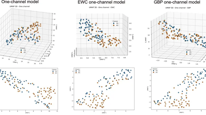
Picture every blood test as a dot on a large map. Plot thousands of those dots and they won't scatter at random: they line up along two main roads. One road separates men from women, the other stretches from youth to old age. Follow these roads and you can see how a person grows up, lives and grows old.
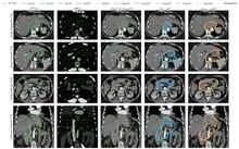
A radiation oncologist looks at an abdominal scan and mentally outlines the area to be irradiated. That area is not visible as a separate organ: its boundaries are set by protocol, not by tissue. So the doctor needs a map of the surroundings to avoid hitting healthy structures.
The nnU-Net model starts with the same task.
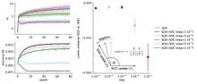
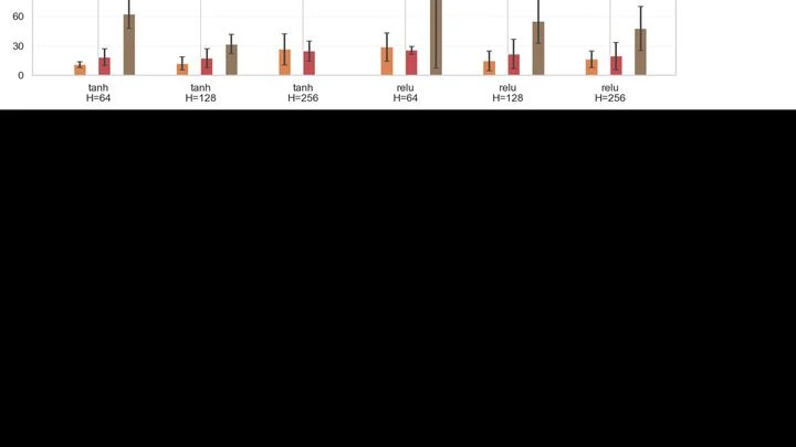
In a nine-story building with a thousand rooms on each floor, some people whisper and others shout. Neighbors pass the message along, but if everyone speaks too softly, the message fades by the third floor. If too loudly, by the top floor it becomes an unintelligible roar. Between these extremes, there is a regime where a whisper makes it all the way through without distortion.
A radiologist looks at an abdominal scan and sees not just organs but their spatial relationships: which kidney is larger, where the tumor lies relative to a vessel. It's like reading a map, where it's not enough to know where a city usually stands; you also need to measure the distance between two points on the ground.
Every heartbeat sends a wave through the cerebral arteries that not only pushes blood but also briefly inflates the vessel. The arterial wall, like a tightly stretched rubber tube, passes that motion to the neighboring brain tissue, and the brain pulses ever so slightly in time with the heartbeat.
In a house where different families live, some age faster, others slower. The same goes for the brain: neurons of different types age at different rates. Some cells vanish by middle age, others hold out into deep old age. Why this happens remained a mystery for a long time.
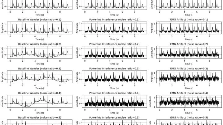
An electrocardiogram is like a recording of a conversation where the heart whispers and a construction site roars in the background. Atrial fibrillation, where the atria quiver instead of contracting, changes that whisper almost imperceptibly.
A protein engineer is like a chef trying to make a dish that is tasty, healthy, and cheap all at once. Change one ingredient, and you risk ruining another. In the past, computer assistants could only evaluate protein properties one at a time, missing how they are connected.
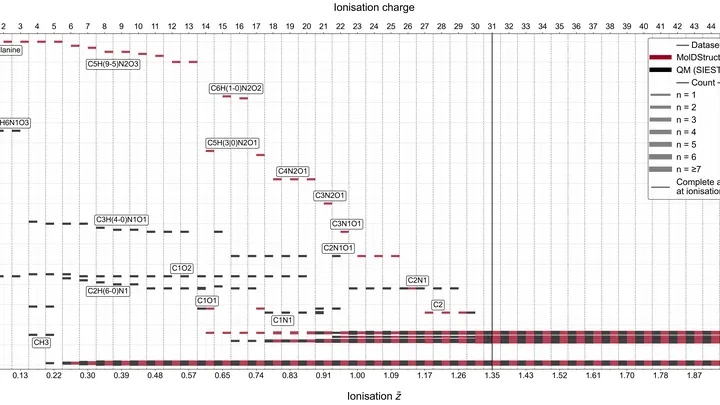
A tennis ball hits a sandcastle. The castle crumbles, but by where and how far the grains of sand fly, you can guess the castle's shape. That's roughly how the method of exploding proteins with X-rays works. The laser beam is so powerful that the protein molecule loses electrons and flies apart into charged fragments, like the castle under the ball's impact.
A single DNA molecule, squeezed into a tiny metal slit, trembles and flickers under the laser. Its signal is not a clear string of letters but a chaotic mix of flashes and shadows. Deciphering this noise is like trying to make out a whisper in a crowded room.
The new neural network works like an attentive interpreter.
Inside a piece of aluminum, atoms are arranged in neat rows, like bricks in a wall. But sometimes a row ends before reaching the edge, and a "step" appears in the crystal, which physicists call a dislocation. These defects determine how strong, ductile, and how a metal behaves under load.
A camera watches a person's face and hands, and from barely perceptible changes in skin temperature it understands: they are struggling right now. Body heat is like invisible ink that shows up under stress. Blood rushes to some areas and drains from others, creating a unique pattern for each person. The challenge is to find the spots and seconds in that thermal signature that betray anxiety.
A medical record is like a long diary: doctor visits, diagnoses, prescriptions, lab results. A human doctor reads it cover to cover to understand what's going on with the patient. Now, a machine reads this diary, and does it in seconds.
The developers created a model called Foresight-E. It was trained on de-identified records of about 61 million people: that's nearly the entire population of England.
Hunting for a new material is like cooking without a recipe: you try, try again, and often throw things out. Previously, scientists manually flipped through papers and tested compositions one by one. Now there's a "robot chef" that reads the literature itself, mixes components, and checks what came out.
At a landfill on the outskirts of town, the wind shifts direction, and a couple of hours later the nearest village smells of rotten eggs. That's hydrogen sulfide — a gas produced when waste decomposes, and in large doses it's hazardous to health. Usually, authorities only learn about the problem after residents have already breathed it in and called to complain.
The instrument shines an infrared beam at a sample, and molecules respond with their own absorption pattern, like bells of different tones. This pattern, the spectrum, holds information about the chemical composition. But to read it, a specialist usually consults a library of reference records, which is slow and not always possible.
The instrument shines an infrared beam at a sample, and molecules respond with their own absorption pattern, like bells of different tones. This pattern, the spectrum, holds information about the chemical composition. But to read it, a specialist usually consults a library of reference records, which is slow and not always possible.
A tumor in a scan is like a tiny bright dot on a vast dark canvas. That dot is what the doctor needs, not a perfectly smooth background. But when a neural network learns to turn a regular CT scan into something resembling a PET scan, it often tries to make everything look nice, and ends up smearing that dot.
A low-dose X-ray image looks like a view through a fogged-up window: outlines are there, but details drown in a hazy veil. Doctors need such images during radiation therapy — to aim the beam precisely at the tumor and spare healthy tissue. But saving on radiation comes at the cost of noise, streaks, and artifacts that obscure what matters.
A doctor spends years learning to recognize diseases from brain scans. First, they excel at spotting signs of aging, then they master tumors, and suddenly they start confusing what they knew before. Many neural networks behave much the same way: new knowledge overwrites the old.
If you pour honey into a colander with a hundred small holes, it will drip in thin streams, but each stream will swirl around the edge of the hole. Engineers need to understand such swirls: they affect how quickly water filters, how oil comes out of a reservoir, or how a drug seeps through tissues.
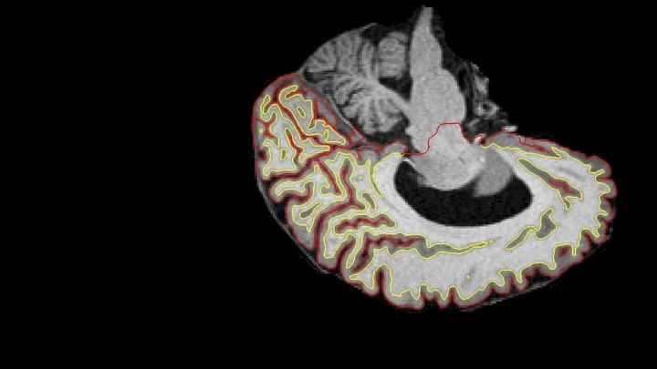
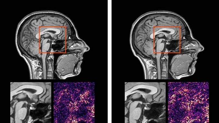
Two scans of the same brain taken at different times are like photographs of the same house before and after renovation. To see what changed, you overlay them and look at the differences. But an MRI scanner is not a perfect camera: it adds its own distortions, as if the photo were taken through slightly warped glass.
A doctor spends years learning to recognize diseases from brain scans. First, they excel at spotting signs of aging, then they master tumors, and suddenly they start confusing what they knew before. Many neural networks behave much the same way: new knowledge overwrites the old.
During surgery, an anesthesiologist watches a screen where pressure and pulse curves run. But behind these lines hide several other quantities: how much the vessels constrict, how fast the blood flows, how elastic the artery walls are. Precise instruments for measuring them are only used in the most severe cases because it's risky — a needle in the artery, a catheter in the heart.
A flashlight shines through frosted glass: the rays bend, lose brightness, and take different paths. That's roughly how high-intensity ultrasound passes through the skull to the brain. For years, calculating this path required bulky computer models that 'chew' on data for hours.
Holding in mind the exact position of a pen as it's slowly moved is a routine task for the brain. For artificial neural networks, it's nearly impossible. Standard recurrent networks like GRU and LSTM tend to break continuous motion into rigid steps: the pen doesn't glide smoothly but jumps from point to point.
A glucose meter that doesn't prick your finger but just looks at your wrist: that's the dream for millions of people with diabetes. It's the promise of algorithms that extract glucose levels from the ordinary pulse signal, the photoplethysmogram (the same one that measures heart rate in smartwatches).
An X-ray machine takes a scan, and the doctor sees only numbers — bone density, fat percentage. But the image holds far more. A team of researchers created a model called LeDXA that reads these scans like a map of hidden bodily signals.
The model was trained on 11,540 scans from the Human Phenotype Project with no labels at all.
A magnetoencephalograph, a helmet-like device with hundreds of sensors, picks up the weak magnetic fields generated by active neurons. When a person listens to speech, these signals contain imprints of the heard words. But extracting them is tricky: signals from thousands of sources merge into a tangled mess.
Predicting the yield of a chemical reaction is a bit like trying to guess the harvest knowing only the soil composition. Traditionally, chemists have relied on long columns of numbers, quantum descriptors, that describe electron energies but say nothing about molecular shape.
The proton beam is like a sniper's bullet: it delivers most of its energy at a precisely set point, barely touching tissues on the way in. But over 4–6 weeks of head and neck tumor treatment, the target shifts: the patient loses weight, the tumor shrinks, posture changes. The sniper aimed at one spot, but the target has already moved.
An array of radio telescopes is like a camera missing several lenses. Ever since Karl Jansky picked up the radio hiss of the Milky Way, astronomers have been trying to piece together a whole picture from fragmentary signals. But the antennas are spaced apart, and the mathematically “developed” image — the dirty map — ends up smeared.
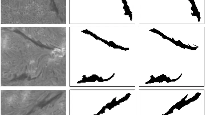
Each morning, dozens of telescopes swing toward the Sun. Their cameras catch a narrow band of red light — the Hα line, discovered by Johann Balmer. The images reveal dark, whimsically curved threads. They look like stitches in a giant tapestry sewn through the blazing corona.
An eye prosthesis sends the brain not a photograph but a sparse pattern of bright dots — phosphenes. To make out a cup or keys in this flickering noise, a person has to move their head like a flashlight in the dark, piecing together the object's outline bit by bit. It's a slow process, like assembling a puzzle where most pieces have been thrown away.
A protein slides along the DNA strand like a key along a keyring, searching for its lock. One wrong turn and a gene stays switched off. To know where a protein will grip firmly, you need to measure the strength of that grip — the binding energy.
By 2025, large language models like GPT are no longer just chatting: they're helping scientists run experiments. But the same tool could fall into the wrong hands. A team of researchers built a digital proving ground to find out.
Every time a child with cerebral palsy is about to move their leg, a distinctive electrical blip appears in the brain — a slow voltage shift. Scientists call it a movement-related potential. A brain-computer interface (a system that reads intentions from brain signals) can pick up this blip and turn it into a command for a training device.
To capture every detail of a statue's shape, you could break it into millions of tiny cubes and describe each one — slow, heavy, and your computer's memory would groan at the seams. That's how 3D medical scans are usually handled: a CT or MRI is a huge array of such cubes (voxels), and to create or improve an image, the machine has to plow through all of them.
The photo enhancement algorithm is like a navigator that step by step leads a blurry picture toward a sharp view. It removes noise and distortions, moving pixel by pixel along a familiar map. But if the image was taken in unfamiliar conditions (say, with unusual blur) the map doesn't match the terrain.