Glowing Clocks Inside the Body express
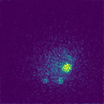
A PET scanner catches flashes of light that occur when a radioactive tracer introduced into the body meets electrons in tissues. Usually, the device merely notes where a flash occurred. But between the birth of the tracer and the flash, there is a pause — tiny, in billionths of a second. During this pause, positronium forms and decays: a short-lived pair consisting of an electron and its antiparticle, the positron. The lifetime of such a pair depends on its environment: in dense surroundings it fades faster, in loose surroundings slower. So if you measure this pause at every point, you get a map not just of “where” but of “in what chemical environment.”
The challenge lies in the volume of data. To build a volumetric map, one must account for millions of possible detector-time combinations, but the instrument actually registers only a small fraction of them. The authors of the study proposed not storing all conceivable variants in the computer’s memory, but keeping only those that actually occurred in the measurements. This makes the model lightweight without losing accuracy: the Poisson statistics (the law of rare events) is preserved.
Then a Bayesian approach comes into play: for each voxel (volumetric pixel), the decay rate is estimated using a formula that updates with each new event, like a score in a game where each point is a registered decay. The formula is closed-form, meaning the answer is obtained directly, without lengthy iterations. On a computer with an ordinary processor, a map of 234,375 voxels was built in about three seconds — versus over a minute for the previous method.
The speed of the calculation opens the door to the clinic: a physician could obtain not only an anatomical image but also a map of tissue chemical activity while the patient is still on the table. This means seeing not just a tumor but also its “character”—how aggressive the environment around it is.
Positronium was predicted long before its discovery: in 1934, Croatian physicist Stjepan Mohorovičić (son of the famous seismologist Andrija Mohorovičić) hypothesized the existence of a bound electron-positron system, but it was only experimentally detected in 1951.
In the series “Star Trek,” the medical tricorder instantly scans tissue biochemistry — positronium lifetime imaging takes a step toward such diagnostics, though for now on the scale of the whole body rather than a handheld device.