How to Spot Details in a Blurry Image express
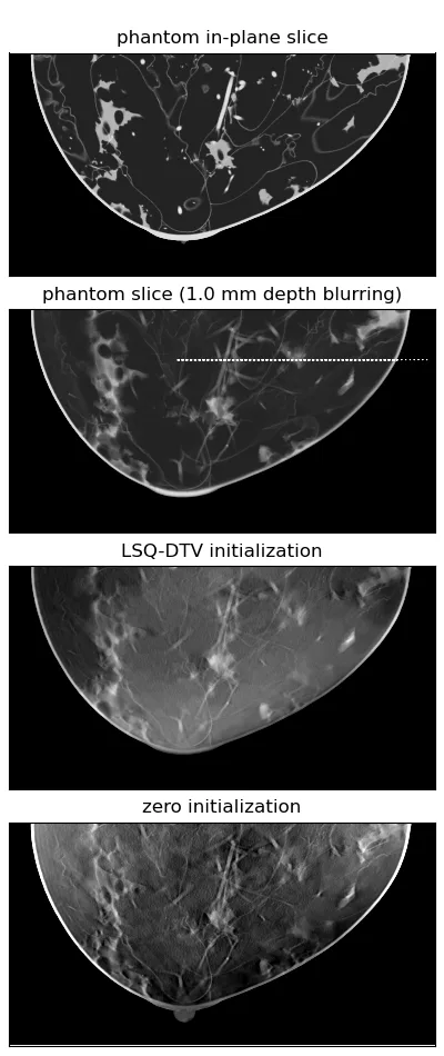
The X-ray machine moves around the breast in an arc, taking several images from different angles. From these flat frames, the computer assembles a 3D picture, like putting together a mosaic from pieces. But if the mosaic pieces are crumpled or smudged, the picture turns out blurry. In tomography, the role of "crumples" is played by noise and imperfections in the imaging.
To make the 3D picture sharp, the reconstruction algorithm needs to be tuned. In the past, tuning felt like cracking a safe: you turn one parameter, then another, and repeat many times. The authors found a simpler way. They showed that for tasks where the image is composed of multiple "layers" with different transformations, a simple rule for choosing the tuning step suffices. No need to try thousands of options — the algorithm adjusts itself.
It's like focusing a camera not by trial and error, but by a simple formula, and getting a sharp shot right away. For patients, this means more accurate diagnosis without extra repeat exams.
Digital breast tomosynthesis takes about 15–25 images in a single pass, while the radiation dose remains comparable to that of a regular mammogram.