method

digital twin healthcare

Using computational models to simulate and predict patient anatomy and treatment outcomes.
38 related articles

Description

A digital twin in healthcare is a computational model that mirrors a patient's anatomy, physiology, or even a specific organ in real time. It integrates data from medical imaging, sensors, and historical records to simulate how a disease progresses or how a treatment might unfold. The goal is to move from reactive medicine to predictive and personalized care, where doctors can test interventions on a virtual copy before applying them to the real patient.

History

The concept of digital twins originated in engineering, notably with NASA's Apollo program in the 1960s, but its application to healthcare gained traction in the 2010s with advances in medical imaging and AI.

How it works

The twin is built from patient-specific data, such as MRI scans, CT images, or continuous monitoring signals. Machine learning algorithms and physics-based simulations then create a dynamic model that updates as new data arrives. For example, a digital twin of a tumor can simulate how it responds to electric fields or drugs, while a twin of the heart can predict arrhythmias or the outcome of surgery. The model is constantly refined by comparing its predictions with actual outcomes, improving its accuracy over time.

In practice

Digital twins are used to plan complex surgeries, optimize radiation therapy, and simulate the spread of epidemics within a city, as well as to train imaging systems like CT scanners.

Fun fact

Digital twins of the brain are being used to select personalized treatments for epilepsy and depression, simulating the effect of electrical stimulation before it is applied.
Concept graph →

Related articles · 38

Optics That Fix Themselves
An orchestra of a hundred musicians plays without a conductor. Every instrument is tuned perfectly, but one violin goes slightly off, and the harmony falls apart.
q-bio.CB2026-09-033 minarXiv:2609.03691
How Tissue Tears: A New Topological Operation in the Vertex Model
A sheet of paper tears where tension exceeds strength. Epithelium, the cell sheet lining our organs, behaves similarly, but until recently models could not show how tears appear in it. The classical vertex model describes tissue as a network of polygons tightly packed together and forbids cells from detaching.
A Street Map for the Radiation Guide
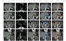
A radiation oncologist looks at an abdominal scan and mentally outlines the area to be irradiated. That area is not visible as a separate organ: its boundaries are set by protocol, not by tissue. So the doctor needs a map of the surroundings to avoid hitting healthy structures. The nnU-Net model starts with the same task.
q-bio.PE2026-08-141 minarXiv:2608.13995express
Epidemics Breathe in Tandem
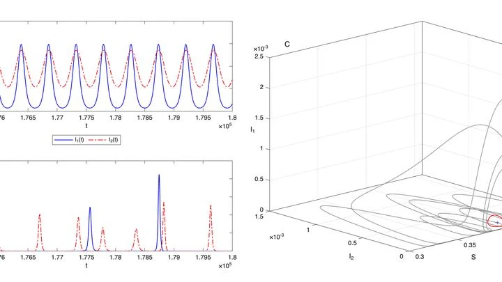
Every winter, hospitals fill up, and by summer they seem to empty. Epidemics breathe like the tide. But why do waves of disease keep rolling in, even without new mutations? Mathematicians found an answer in a tug-of-war between two strains. Within a single epidemic, two virus variants often coexist.
Image Processing2026-08-082 minarXiv:2608.08173express
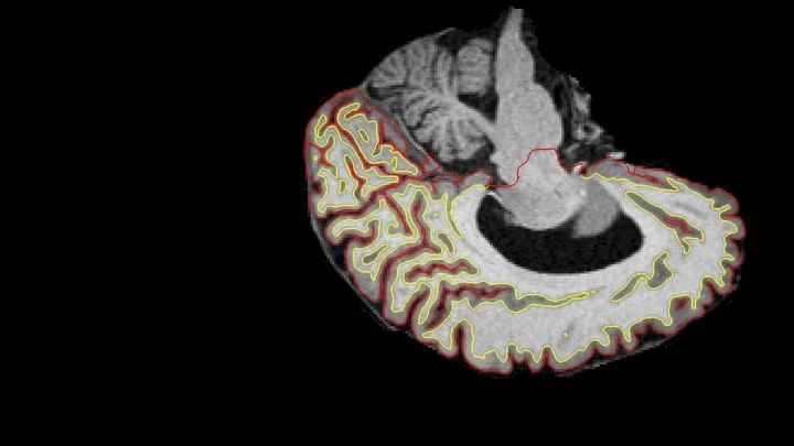
Separating Brain Scans: Where Disease Ends and Distortion Begins
Two scans of the same brain taken at different times are like photographs of the same house before and after renovation. To see what changed, you overlay them and look at the differences. But an MRI scanner is not a perfect camera: it adds its own distortions, as if the photo were taken through slightly warped glass.
Medical Physics2026-08-032 minarXiv:2608.02889express
Water Sees Radiation
Radiation therapy is a precision strike on a tumor, but every shot must be calibrated to within a millimeter and a fraction of a second. The problem is that existing dosimeters are either bulky, or they distort the beam themselves, or they require pauses for readout.
Medical Physics2026-08-012 minarXiv:2608.00831express
Digital Twin Predicts Patient Anatomy
The proton beam is like a sniper's bullet: it delivers most of its energy at a precisely set point, barely touching tissues on the way in. But over 4–6 weeks of head and neck tumor treatment, the target shifts: the patient loses weight, the tumor shrinks, posture changes. The sniper aimed at one spot, but the target has already moved.
eess.SY2026-07-151 minarXiv:2607.13766express
Digital Twin: How People's Invisible Behavior Changes Plans
In any city project, residents' behavior is a invisible force that determines success. One skeptical neighbor can slow down solar panel installation across an entire block: their caution triggers a rise in disorder.
Image Processing2026-07-151 minarXiv:2607.13812express
A Fresh Perspective on 3D Medicine
The data from a single CT scan is comparable to several feature-length films. TCAM-Diff bypasses this bottleneck: the program compresses the 3D scene into three shadow projections — like a sculptor reconstructing a solid statue from silhouettes on three walls.
eess.SY2026-07-151 minarXiv:2607.13914express
Epidemic Under Control: A Recipe for Systemic Approach
When a pipe bursts in the basement, a plumber acts step by step: locates the leak, measures the pressure, determines the cause, patches it up, and checks if it's dry. The same recipe — the standard model DMAIC — is being proposed against epidemics. First, they determine the scope — how many hospital beds are occupied, which industries are sagging.
Image Processing2026-07-141 minarXiv:2607.12586express
Water's Geometry Improves Medical Scans
Medical scans are maps of tissue density. The brightness of each pixel reveals what’s there, much like photometry measures light intensity. But conventional algorithms analyze pixels in isolation, producing jagged organ boundaries. A new approach compels the program to prioritize smoothness.
Medical Physics2026-07-131 minarXiv:2607.11290express
Threads in a CT Scanner: Algorithm Untangles Catheters in Seconds
In brachytherapy, thin catheters are inserted into tumors. On CT scans, they look like tangled glowing threads. Previously, doctors would manually outline each one, spending up to an hour. The new algorithm learns from synthetic scans — drawing metal strips in an empty volume, like threads without a body.
cs.OH2026-07-013 minarXiv:2607.19912
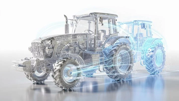
A Tractor’s Digital Shadow: An Exact Copy in Motion
The tractor traces a perfect circle on the stubble. At that same moment, its digital shadow, an exact virtual copy, repeats the maneuver on a computer screen, diverging from the original by only a few percent.
Who Heals Better: General AI or a Narrow Specialist
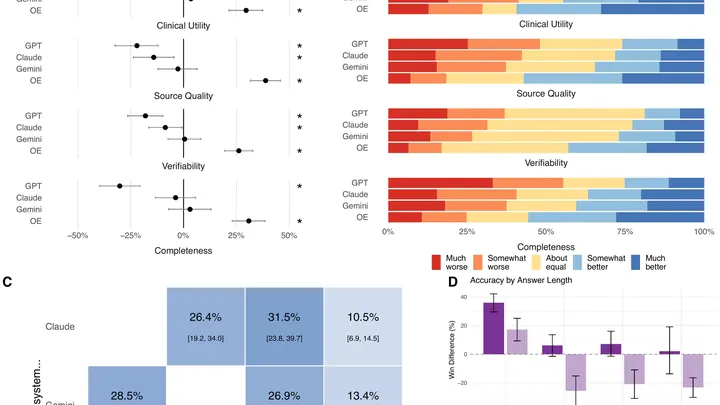
Every week, doctors ask AI assistants millions of questions at the bedside. But these tools are usually tested on exam-style problems, not on what clinicians actually ask in practice. A team of researchers set out to fix that. They collected 620 real questions from physicians across 30 specialties — from cardiology to oncology.
Disordered Systems2026-06-131 minarXiv:2606.15344express
Light Condensate Learns to Draw Digits
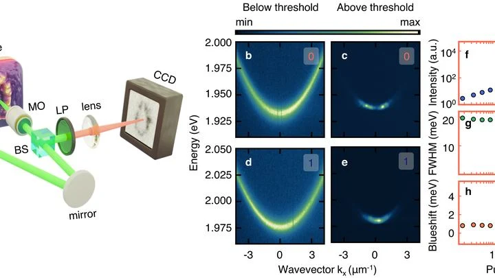
Generative neural networks that create images typically rely on digital randomness. But a team of researchers built a system where randomness is born not in code, but in a tiny optical cavity filled with an organic dye.
q-bio.QM2026-06-092 minarXiv:2606.11264express
A Digital Twin of Health as an Orchestra of Organs
A grand symphony orchestra plays, but each group of instruments follows its own score without hearing the others. The violins carry the melody of the heart, the cellos the rhythm of the liver, and the woodwinds the pulse of the brain. It sounds disjointed, even though each part is flawless on its own.
Machine Learning2026-05-302 minarXiv:2606.00892express
How a Digital Twin Saves a Brain in Minutes
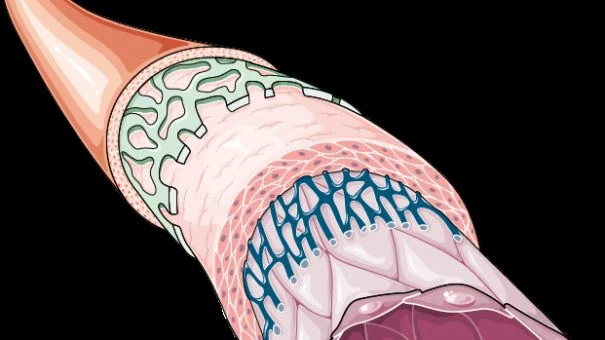
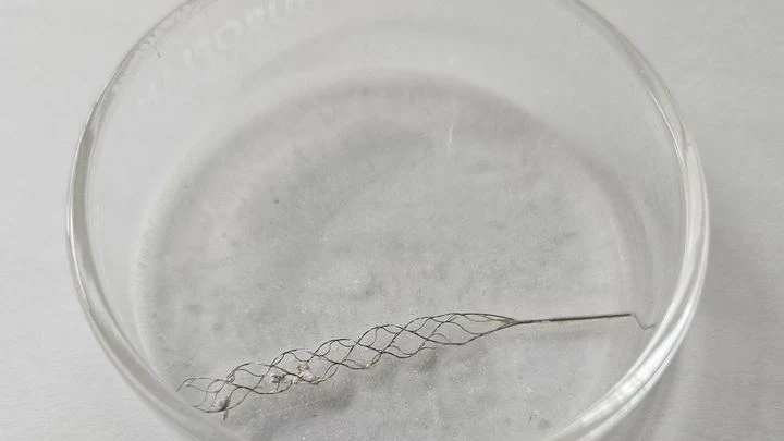
A plumber clears a clog in a narrow, winding pipe — by feel, without seeing inside. That's roughly how a surgeon feels during mechanical thrombectomy, the operation to remove a blood clot from a brain vessel. Time is short, and every move can either save a life or damage the vessel wall.
Medical Physics2026-05-242 minarXiv:2605.24815express
How to Make MRI See a Beating Heart
A photographer shoots a runner with a long exposure and gets a blurry silhouette. That's roughly how a standard MRI scanner "sees" moving organs: the heart contracts, the lungs breathe, and the image smears. To avoid this, patients are asked to freeze and hold their breath. But for a child or someone short of breath, that's nearly impossible. The method called GRASP tackles the problem differently.
Machine Learning2026-05-162 minarXiv:2605.16860express
Sugar Under Control: A Model with a Digital Twin
A person with type 1 diabetes drives a car in thick fog, seeing only a few meters ahead. Blood sugar can plummet or spike, and the insulin pump must react in advance. A four-hour forecast is an attempt to glimpse the road's turn through the haze.
Medical Physics2026-05-093 minarXiv:2605.12544express
A Vascular Twin Made of Digits
The vessels inside the skull resemble a mountain road's hairpin turns — sharp bends, narrowings, unexpected curves. When a traffic jam forms on such a serpentine, blood stops reaching the brain properly. To predict trouble, doctors need to see the entire flow: where it accelerates, where it swirls into eddies. The full picture comes from a special MRI, like slow-motion cinematography of fluid.
Machine Learning2026-04-252 minarXiv:2604.23368express
How a neural network reads a medical history
A doctor holds a stack of test results taken at different times. Each sheet on its own is just a snapshot of a single day, but what you want is a whole movie of how the disease unfolded. A neural network called Tempo learns to glue these scattered frames into a smooth reel.
Machine Learning2026-04-251 minarXiv:2604.23465express
Digital Twin Instead of a Control Group
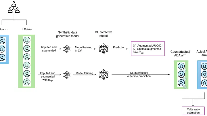
The old paint on one board has already dried, and the new coat hasn't been applied yet, but you need to know which one protects better against rain. Usually, you'd paint two boards and wait a year. The researchers did something different: they taught a computer to predict, from old photographs, how the same board would look under a different paint. No real experiment needed.
Optics2026-04-241 minarXiv:2604.22660express
Light Instead of Wires: Photonic Chip Computes Faster
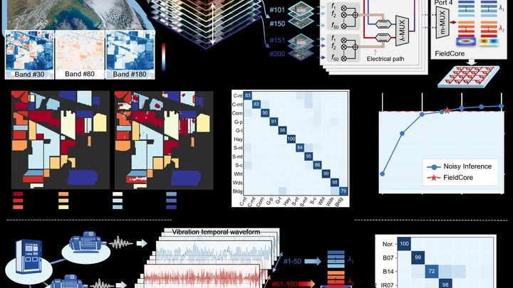
A typical computer shuttles electricity along copper traces, and every operation is a queue: first one thing, then another. Light works differently. In a single beam, you can mix dozens of signals without them interfering, like different voices in a choir, singing at once but each heard distinctly. The new FieldCore chip uses five such "voices" at once.
math.OC2026-04-241 minarXiv:2604.23063express
How to Spot Details in a Blurry Image
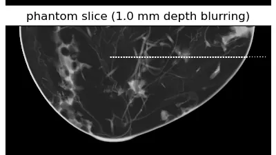
The X-ray machine moves around the breast in an arc, taking several images from different angles. From these flat frames, the computer assembles a 3D picture, like putting together a mosaic from pieces. But if the mosaic pieces are crumpled or smudged, the picture turns out blurry. In tomography, the role of "crumples" is played by noise and imperfections in the imaging.
Medical Physics2026-04-161 minarXiv:2604.14494express
A Ring That Feels Your Pulse
Your finger is a living sensor: beneath the skin, arteries pulse in time with your heart. Traditional blood pressure cuffs squeeze your arm and give a single number, but this ring uses eight electrodes to pass a weak current through the tissue and map blood flow.
A Virtual Brain Chooses the Right Treatment
The brain of a person with Parkinson's disease works like an orchestra where the conductor either freezes or waves the baton off-beat. Medications don't help everyone, so doctors turn to stimulation — weak electrical pulses that restore the rhythm.
physics.acc-ph2026-03-301 minarXiv:2603.28549express
Digital Twin Teaches a Laser to See
A laser strikes a tiny glass grating, and electrons passing through it are accelerated. You can't see inside, so you judge what's happening by the energy of the electrons after acceleration, like judging the shape of a stone by the splash it makes. Scientists built a digital twin: a computer model that mimics the behavior of the real accelerator.
Medical Physics2026-03-271 minarXiv:2603.27003express
Heart by Contour: How AI Builds 3D Shape
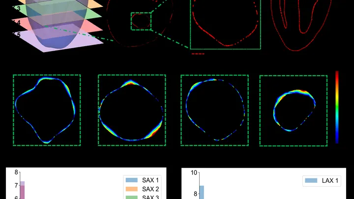
The heart is not a perfect sphere; its shape changes with every beat. To build an accurate 3D copy for a computer model, doctors typically take dozens of MRI slices. But that's like assembling a watermelon from thin slices: the fewer slices, the more guesswork. A new approach teaches a neural network to understand the heart's shape from sparse contours.
cs.CE2026-03-202 minarXiv:2603.19829express
The Brain as Clay: Why It Shrinks in Alzheimer's Disease
Alzheimer's disease begins quietly: deep in the brain, two proteins, tau and amyloid-beta, start to accumulate. They behave like weed seeds: one clump that appears causes neighboring cells to produce similar clumps. Gradually, the proteins spread along nerve fibers, like water through the capillaries of a paper towel, and invade new areas.
Medical Physics2026-03-152 minarXiv:2603.14562express
Tumor Memory: How Fractional Order Changes the Prognosis
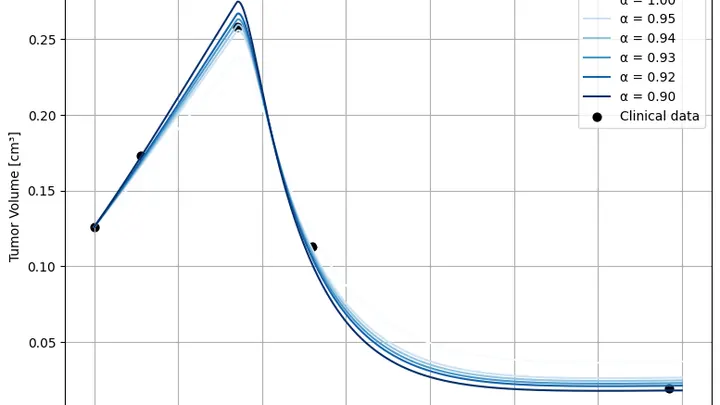
A tumor doesn't grow like a tree; it grows like a city: today's skyline is shaped by yesterday's construction and the fires of the day before. Standard tumor growth equations don't account for this: they only look at the current moment, as if the city were rebuilt from scratch every day. The authors added "memory" to the model: now the equation remembers what came before, and that changes the forecast.
Signal Processing2026-02-281 minarXiv:2603.16891express
AI Reads Cardiograms Around the Clock
An electrocardiogram is like a long strip of paper on which the heart leaves its signature. A doctor studies it to catch rhythm disturbances, but when the recording lasts for days, the strip stretches for miles. The eye tires, attention wanders, and rare but dangerous irregularities can slip by. AI-HEART works like a tireless assistant that scans the entire strip without a break.
Medical Physics2026-02-162 minarXiv:2602.14803express
How the Sound of Light Speeds Up Body Imaging
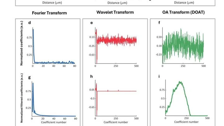
A laser beam hits the skin, and tissues respond with a barely audible ultrasonic echo. From that echo, doctors can see blood vessels and tumors without making a single incision. But before the picture appears on the screen, the signal has to be cleaned of noise, just as a cook skims the foam off a broth to make the soup clear.
Medical Physics2026-02-131 minarXiv:2602.12917express
Ultrasound Keeps an Eye on the Spine
Your back hurts, and the doctor asks you to bend forward and backward to see how your vertebrae move. Usually, this means a series of X-rays: they show motion, but only in a plane, like a shadow on a wall. And volumetric methods like CT scans can't keep up with movement. Engineers proposed a different approach: a robot sweeps an ultrasound probe over your back while you bend.
Optics2026-02-041 minarXiv:2602.04618express
Counting Blood Cells Without Lenses
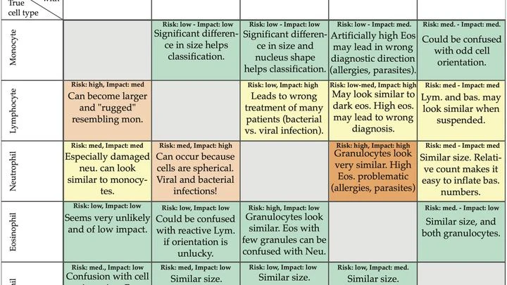
A drop of blood lands on a tiny sensor, and within a minute the doctor knows how many different defenders are in it. That's the future of blood testing, brought closer by Danish engineers. Instead of a regular microscope with lenses, they built a lensless device. A cell flows over the sensor, which records a pattern of dark and light rings — a hologram.
Fluid Dynamics2026-01-272 minarXiv:2601.19994express
Predicting Blood Damage in Pumps
In a heart-lung machine, blood flows through narrow tubing and past rapidly spinning pump blades. Sometimes red blood cells can't take it: their membranes rupture, and hemoglobin spills into the plasma. For a patient, this is like pouring sand into a car's fuel system — small damages accumulate and wreck the whole mechanism.
Medical Physics2026-01-221 minarXiv:2601.15947express
A Camera That Can See Tissues Breathe
Cells breathe. They need oxygen, and they take it from the blood flowing through the tiniest vessels. If blood flow is weak or oxygen is scarce, cells start to suffocate. Doctors need to see this as early as possible. But how do you look inside living tissue without cutting it open? The new device does it in two ways at once.
Medical Physics2026-01-141 minarXiv:2601.09235express
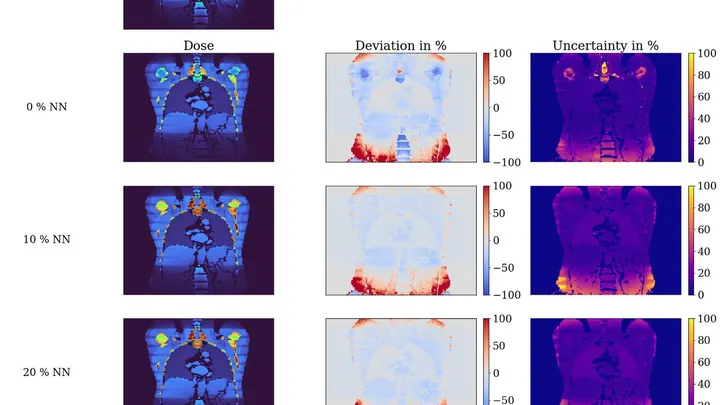
Digital Twins Teach the CT Scanner
Computed tomography is like an X-ray that takes hundreds of images from different angles and stitches them into a 3D picture of the body. But each image delivers a small dose of radiation, and doctors need to know how much each organ receives. Currently, this is often estimated roughly using average tables rather than for the specific patient.
Optics2026-01-022 minarXiv:2601.00552express
X-ray Without a Camera: How to Film a Spinning Engine
An X-ray image is like a photograph taken in the dark with a very short flash: anything that moves comes out blurred. Conventional X-ray detectors are large arrays of millions of pixels, and by the time they read out one frame, the next is already missed. For a spinning turbine or a beating heart, such sluggishness is a death sentence. A team of researchers sidestepped this problem by ditching the array altogether.