Protein Explodes Under X-rays express
A tennis ball hits a sandcastle. The castle crumbles, but by where and how far the grains of sand fly, you can guess the castle's shape. That's roughly how the method of exploding proteins with X-rays works. The laser beam is so powerful that the protein molecule loses electrons and flies apart into charged fragments, like the castle under the ball's impact.
The challenge is that calculating such an explosion for a protein made of thousands of atoms is beyond ordinary quantum methods. The new program MolDStruct combines two approaches: stochastic ionization processes (like rolling dice) and classical physics of atomic motion (like calculating billiard ball trajectories). First, the program "rolls the dice"—it decides which atom loses an electron under the beam. Then it calculates how the remaining charged atoms repel each other and fly apart.
Next, MolDStruct was tested on a protein made of 16 amino acids. A protein can fold into different shapes, like a rope twisted in different ways. The program showed that these shapes can be distinguished from each other by the map of scattered fragments. This means that the explosion of a protein is not just destruction, but a way to peer into its structure.
The method is still young, but it's already clear: the X-ray beam, which used to only damage samples, now helps us see the invisible. Just as the scattered grains of sand can reveal the castle's shape, so can the protein's fragments reveal its structure.
Original abstract
as written by the authors arXiv:2608.17460v1
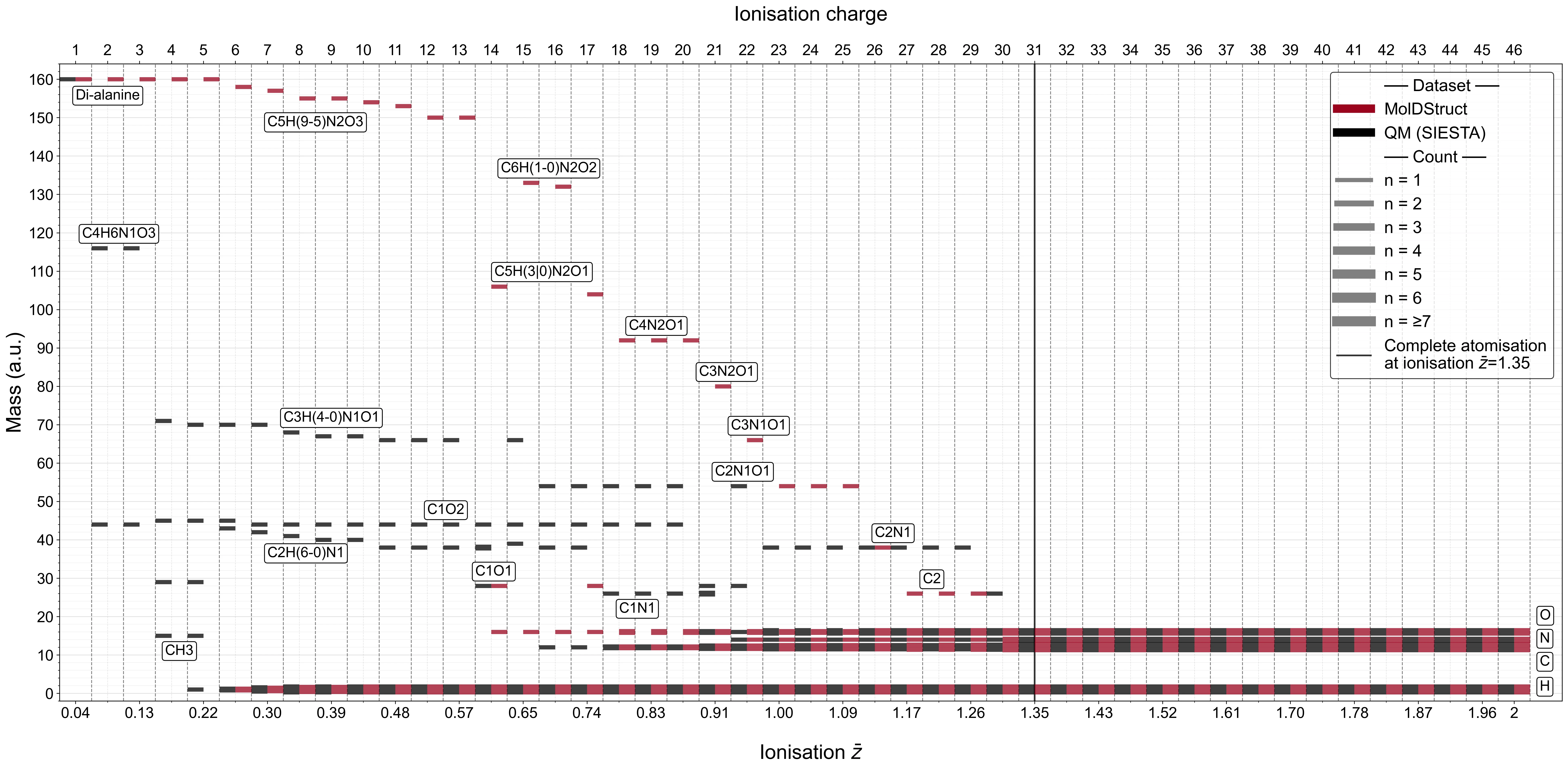
Single Particle Imaging with intense X-ray free-electron laser pulses requires modelling of the resulting ionisation and Coulomb explosion dynamics of biomolecules to optimise experimental parameters and enable correct structural reconstruction, yet simulating the complete dynamics at protein scale is beyond the reach of quantum-mechanical methods. To address this, we developed \moldstruct, a hybrid Monte Carlo/Molecular Dynamics code built on GROMACS that couples high intense X-ray ionisation dynamics modelled through a Monte Carlo module with classical Molecular Dynamics for atomic propagation. Benchmarked against quantum mechanical calculations for di-alanine, MolDStruct agrees in fragmentation patterns above a mean charge per atom of $$\bar{z} \approx 1.35$$. Compared with Coulomb explosion imaging experimental data for 2-iodopyridine, simulated momentum distributions reproduce the experimental Newton plots in fragment direction and fall within the range of experimental absolute momenta, with a moderate overestimation. We further apply MolDStruct to Protein Explosion Imaging, a method that classifies molecular structures from explosion ion maps recorded on a detector, demonstrating via dimensionality reduction that conformers of the 16-residue peptide $$\mathrm{Ala}_16$$ and five chromophore-labelled ubiquitin mutants are distinguishable. These results establish MolDStruct as a practical tool for simulating radiation damage and Coulomb explosion dynamics of biomolecules at protein scale, where quantum-mechanical methods are computationally infeasible.
The X-ray laser is so powerful that it destroys a protein in millionths of a second — faster than the molecule can "feel" the impact.