How to Teach a Program to Draw the Radiation Zone ✛
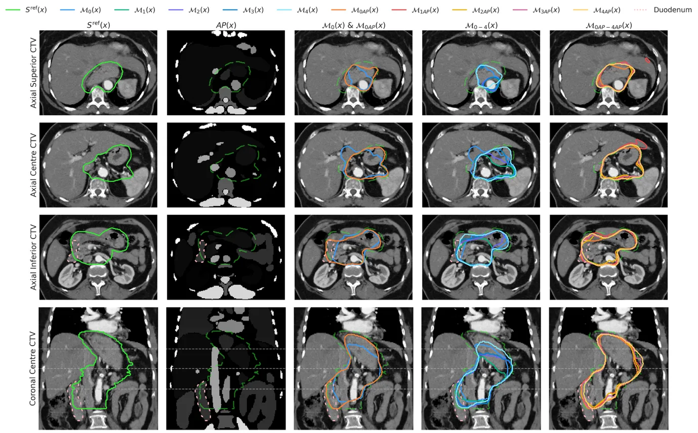
When a doctor treats stomach cancer with radiation, they must precisely mark the area to be irradiated. This area is not visible as a separate organ, so the doctor has to keep the locations of all neighboring organs in mind. The program nnU-Net learns to do the same, but first it needs to be shown examples.
Researchers gave the program a map with all abdominal organs marked. It is like a street map with labeled buildings: the program sees where the stomach is, where the intestine is, and does not stray into foreign territory. They also further trained it on the hardest cases, those where the program itself was not confident in its answer. In this way, it learns from its mistakes.
After such training, the program outlines the boundary almost as well as experienced doctors: its score is 0.87, while doctors' scores range around 0.89. Now the doctor can trust the program with a draft and only check questionable cases. This saves time and helps treat patients.
Training one model took about 22 hours on an NVIDIA Quadro 8000 GPU, while labeling anatomical landmarks for one CT scan took only 6 minutes. Adding this six-minute map saved four rounds of active learning, each of which required manual contour review by two oncologists.