Anatomical Priors and Active Learning Improve Target Delineation in Gastric Cancer Radiotherapy ✛
Context
This work stands somewhat apart among studies on automated contouring in radiotherapy: its closest neighbors focus on tumor segmentation on MRI and CT, dimensionality reduction, and weakly supervised learning. It has been shown that deep networks segment organs and tumors reasonably well, but for the clinical target volume, which is defined not by tissue boundaries but by protocol and anatomical landmarks, ready-made solutions are scarce. In the TOPGEAR trial, this volume is described through multiple abdominal structures, and manual contour review for hundreds of patients is too time-consuming. Hence, a model that proposes a reference contour on its own is needed, with the physician only reviewing suspicious discrepancies.
Methods
We took 100 CT scans from the TOPGEAR trial: the first 10 patients formed the initial training set, 40 simulated sequential data arrival, and 50 were held out as a test set. For each initial case, five radiation oncologists independently contoured the CTV, and their versions were combined using the STAPLE algorithm into a reference contour. The base model was nnU-Net (3D full-resolution configuration), trained with five-fold cross-validation. The anatomical prior was built as follows: TotalSegmentator labeled 117 structures on each scan, the resulting map was normalized and fed as a second channel alongside the CT image. Active learning was simulated with four rounds: in each round, the model processed the next batch of 10 cases, and for fine-tuning, cases were selected where model confidence was below the 25th percentile or the Dice coefficient fell below 0.85. Confidence was assessed by the spread of predictions from five ensemble models in a five-millimeter band around the contour boundary.
Results
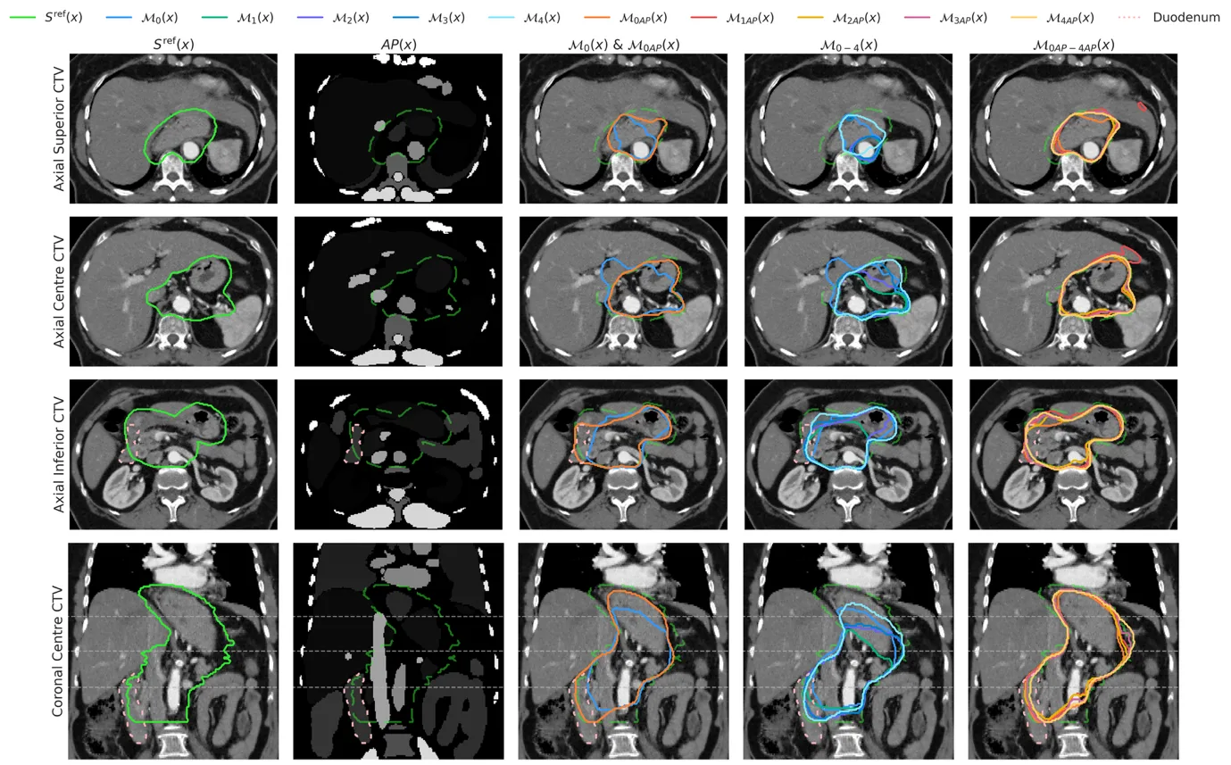
The anatomical prior immediately improved segmentation: the mean Dice coefficient rose from 0.84±0.09 to 0.86±0.05, and inter-patient variability decreased. Active learning without the prior gave a smaller gain: only by the fourth round did accuracy reach 0.86±0.05. Combining both techniques yielded the best result, 0.87±0.03, which closely approached the inter-expert variability of 0.89±0.02 measured on the initial set. Model confidence correlated positively with the Dice coefficient (Pearson coefficient around 0.6–0.7), meaning the model itself knew where it was making mistakes. An example slice with contrast shows how the model with the prior more accurately covered the duodenum and stomach, structures not present in the initial training set.
Implications
This work aligns with neighboring research on medical image segmentation but differs in its target and validation approach: it is the first to show, for a complex clinical volume such as the CTV in gastric cancer, that standardized anatomical maps from the general-purpose TotalSegmentator model yield accuracy gains comparable to four rounds of active learning. Neighboring studies on mediastinal lymph nodes gave conflicting results on the benefit of such priors, whereas here the effect is statistically significant and reproducible on an independent test. This strengthens the case for anatomical priors as a cheap and reliable way to improve segmentation of volumes defined by protocol rather than visible boundaries.
Future development
Active learning could be continued until accuracy plateaus rather than stopping at four rounds. Instead of a voxel-wise structure map, distance maps to key organs could be tried: for anatomy-driven volumes, they might provide smoother spatial context. A separate question is how to transfer this approach to other sites where the CTV is defined differently, such as the prostate bed or the postoperative breast bed. It also remains to be tested how much the gain in Dice coefficient translates into a real reduction in missed protocol deviations during automated contour checking.
Impact
The results are directly applicable in radiation oncology departments that implement automated contour checking for clinical trials. Manufacturers of autocontouring systems could integrate TotalSegmentator anatomical maps into their products. The active learning methodology will benefit any research group building segmentation models as patients accrue in a study.
Next steps
Evaluate the sensitivity and specificity of automated contour checking based on this model compared with manual review. Run additional rounds of active learning to find the saturation point in accuracy. Adapt the approach to other clinical volumes and compare with alternative sources of anatomical priors, such as specialized models for radiotherapy.
Key open problems
In medical segmentation, the question of how to train models on small and heterogeneous datasets without losing generalization remains open. This work shows that anatomical priors and active learning are two practical tools that can be combined, but their effects partially overlap. It remains unclear how to select selection thresholds and uncertainty parameters without tuning on each new dataset. The calibration of model confidence is also an open problem: the correlation with accuracy exists, but whether it is sufficient for clinical decision-making remains to be determined separately.
The knowledge machine's view
This section is addressed to the author of the paper and their colleagues. Neighbouring works were found by meaning-based search across our archive, not by word overlap. We do not review: we show the field around the work.
What the work does
The study integrates anatomical landmark maps from TotalSegmentator into nnU-Net and applies active learning with low-confidence case selection for CTV contouring in gastric cancer within the TOPGEAR trial. Both approaches raised the mean Dice coefficient from 0.84 to 0.87, approaching the inter-expert variability of 0.89. The model demonstrated an ability to estimate its own confidence, which correlated with accuracy.
Where its strength is
The work shows that standardized anatomical maps from the universal TotalSegmentator model yield accuracy gains for a challenging clinical volume, comparable to several rounds of active learning. This paves the way for automating contour review in clinical trials.
Where it could go next
- It might be interesting to see how the anatomical landmark approach combines with architectures where dense connections improve feature utilization: our archive includes an analysis of DenseNet-121 for brain tumor segmentation, where such connections provided an advantage over ResNet-50.based on: Dense Connections vs. Residual Paths: How DenseNet-121 Outperforms ResNet-50 in Brain Cancer Detection arXiv:2505.11735
- Nearby is a study on AI-based contouring of cancer in whole-body scans; transferring ideas from that work to gastric CTV might improve generalization to rare anatomical variants.based on: AI Outlines Cancer in Whole-Body Scans arXiv:2607.03931
- The archive contains research where a neural network learns to detect tumors by combining multiple modalities; such a multimodal approach could be useful for CTV, where not only CT but also other data are important.based on: Two Eyes Are Better Than One: How a Neural Network Learns to See Tumors arXiv:2604.23137
Worth trying
- Test whether replacing TotalSegmentator with a more specialized abdominal segmentation model provides additional accuracy gains.
- Evaluate how active learning with confidence-based selection behaves with more rounds and a smaller initial training set.
- Compare the proposed method with fully automatic contouring that does not involve expert input during the labeling phase.
Nearby works in our archive
- Water's Geometry Improves Medical Scanssimilarity 0.58
- Gene Map Predicts Breast Cancer Outcomessimilarity 0.54
- AI Outlines Cancer in Whole-Body Scanssimilarity 0.54
- Dense Connections vs. Residual Paths: How DenseNet-121 Outperforms ResNet-50 in Brain Cancer Detectionsimilarity 0.54
- Two Eyes Are Better Than One: How a Neural Network Learns to See Tumorssimilarity 0.52
The archive is sparse around this work: the closest review is only 0.58, against a usual 0.64. It looks like few have drilled nearby.
Training one model took about 22 hours on an NVIDIA Quadro 8000 GPU, while labeling anatomical landmarks for one CT scan took only 6 minutes. Adding this six-minute map saved four rounds of active learning, each of which required manual contour review by two oncologists.
Key numbers
- Dice coefficient without improvements: 0.84
- Dice coefficient with anatomical prior and active learning: 0.87
- Inter-expert variability: 0.89
- Number of initial training cases: 10
- Number of active learning rounds: 4