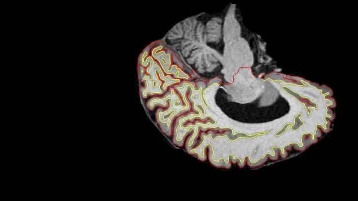
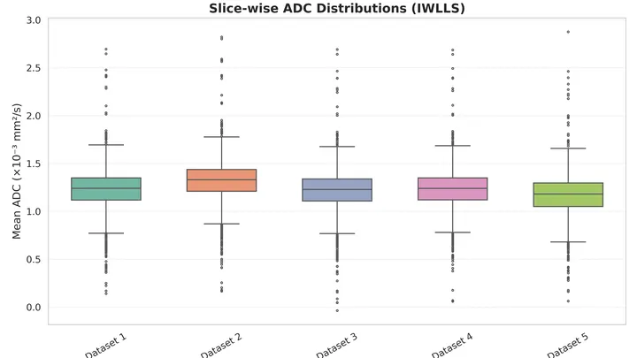
Medical imaging AI refers to machine learning systems, especially deep neural networks, that automatically analyze medical images such as CT scans, MRIs, and ultrasounds to detect abnormalities, segment anatomical structures, and predict clinical outcomes. These systems are becoming indispensable in radiology, where they help clinicians spot tumors, measure disease progression, and even forecast how a patient will respond to treatment. The field matters because it promises to increase diagnostic accuracy, reduce workload, and catch diseases earlier than the human eye alone.
History
The use of computers in medical imaging dates back to the 1960s with computer-aided detection, but modern deep learning approaches emerged after 2012, following breakthroughs in image recognition. The field accelerated rapidly with the availability of large annotated datasets and GPU computing.
How it works
The AI is trained on large datasets of labeled medical images—for example, thousands of CT scans with tumors marked by radiologists. During training, the network learns to recognize patterns and features that correlate with the labels. Once trained, it can process a new scan in seconds, outputting a probability map of where a tumor might be, or a classification of whether the image shows signs of a particular disease. Advanced architectures like convolutional neural networks (CNNs) and transformers are used, often with techniques to handle the high resolution and 3D nature of medical scans.
In practice
AI systems are already deployed in hospitals to flag suspicious lesions in lung CTs, assist in breast cancerscreening, and segment brain tumors for surgical planning, as well as in research to predict treatment responses from imaging data.
Fun fact
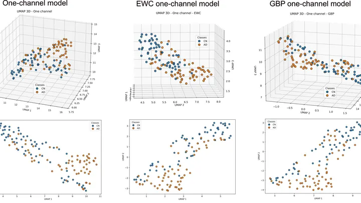
Some AI models can detect subtle changes in medical images that are invisible to the human eye, such as early signs of Alzheimer's disease years before symptoms appear.
An old mattress stops bouncing back: the filling clumps up, and the springs lose their resilience. The ovary changes with age in much the same way, except that instead of filling, stiff connective tissue grows inside it, and the role of springs is played by the cells that support the eggs.
In a hall of mirrors, a person walks up to a curved mirror and sees themselves now as a dwarf, now as a giant. Popular machine learning models behave in much the same way when asked to guess biological age from blood tests or brain scans: they stretch age at the extremes, underestimating it in older people and overestimating it in younger ones.
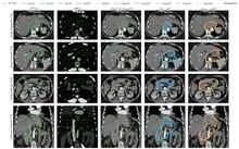
A radiation oncologist looks at an abdominal scan and mentally outlines the area to be irradiated. That area is not visible as a separate organ: its boundaries are set by protocol, not by tissue. So the doctor needs a map of the surroundings to avoid hitting healthy structures.
The nnU-Net model starts with the same task.
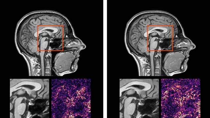
Two scans of the same brain taken at different times are like photographs of the same house before and after renovation. To see what changed, you overlay them and look at the differences. But an MRI scanner is not a perfect camera: it adds its own distortions, as if the photo were taken through slightly warped glass.
A doctor spends years learning to recognize diseases from brain scans. First, they excel at spotting signs of aging, then they master tumors, and suddenly they start confusing what they knew before. Many neural networks behave much the same way: new knowledge overwrites the old.
To capture every detail of a statue's shape, you could break it into millions of tiny cubes and describe each one — slow, heavy, and your computer's memory would groan at the seams. That's how 3D medical scans are usually handled: a CT or MRI is a huge array of such cubes (voxels), and to create or improve an image, the machine has to plow through all of them.
Communicating with a satellite is like trying to hear a whisper in a storm: the signal drowns in noise. Engineers have created an AI algorithm that runs directly on the satellite and, with minimal power consumption, detects distortions more accurately than previous methods.
When a pipe bursts in the basement, a plumber acts step by step: locates the leak, measures the pressure, determines the cause, patches it up, and checks if it's dry. The same recipe — the standard model DMAIC — is being proposed against epidemics.
First, they determine the scope — how many hospital beds are occupied, which industries are sagging.
Ultrasound waves bounce off organs to draw their outlines, but on an ovarian scan it's hard to tell a harmless fluid-filled cyst from a solid cancerous mass. Even specialists draw the boundaries differently, and this directly affects treatment choices.
The OvAi Focus program tackles this by learning from thousands of examples.
A child exploring an unfamiliar apartment crawls from object to object and remembers what is visible from each corner. This is how the brain builds a mental map — an internal model of space. The gradCSCG algorithm works on the same principle: it receives a video stream from a camera and independently draws a map of the room, relying only on the changing images.
Prostate MRI scans often look like photos taken through a dirty window: noise, blurred edges, curvature from field imperfections. Scientists checked what happens if you 'wash' that window: remove the excess, correct the distortions, and accurately align different types of scans.
Building a light chip is like a game where you have to guide a beam through a maze of tiny mirrors and beam splitters. Each element acts like a railroad switch: it either lets light pass straight through or directs it to an adjacent track. The goal is to produce a specific light pattern at the output while using as few 'switches' as possible.
Usually, engineers spend years finding the shortest route for each task.
An electron microscope is a giant pipe organ: instead of pipes (electromagnetic lenses, instead of air) a stream of accelerated electrons. It once merely painted mute frescoes of the atomic world, condemning scientists to grueling decipherment.
The Universe is less than a billion years old, yet at its centers already sit black holes with masses of a billion suns. According to the conventional picture, they grew by voraciously devouring matter, but calculations show that 'feeding' must have been almost continuous, while visible quasar flares last only a few million years. So where does all the accreting fuel go?
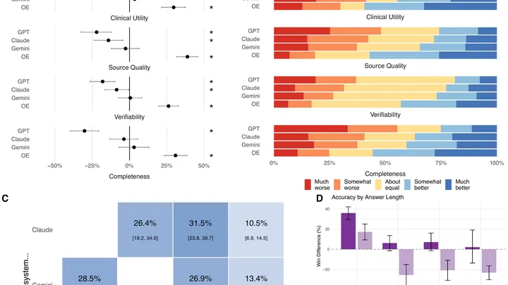
Every week, doctors ask AI assistants millions of questions at the bedside. But these tools are usually tested on exam-style problems, not on what clinicians actually ask in practice. A team of researchers set out to fix that.
They collected 620 real questions from physicians across 30 specialties — from cardiology to oncology.
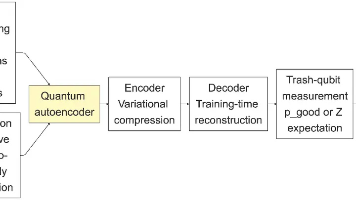
A cook spends years honing the skill of reducing a recipe to three or four essentials and recreating the dish from them. If the ingredients are familiar, the expected meal lands on the plate. A foreign ingredient disrupts compression, and you get something unrecognizable. The Quantum Compressor does the same with medical scans.
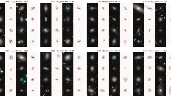
An ordinary photo editor highlights objects in an image. The ZooBot:3D neural network works with distant galaxies like a cartographer drawing a map of an unexplored land.
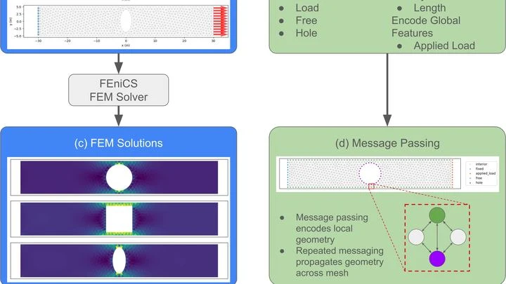
Designing machines is like cooking: you need to know exactly where the 'dough' will tear. Old computer simulations check every point on a part for hours, like measuring the entire pot to make sure the handle doesn't fall off.
A single camera shot stores both a normal picture and a hidden map of how light rippled through the scene. Quantum entanglement makes this possible. Pairs of linked photons are generated: one visits the object, its twin takes a detour. Later, measuring the twin one way pulls out the brightness information.
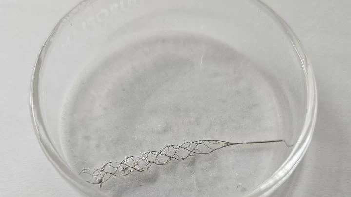
A plumber clears a clog in a narrow, winding pipe — by feel, without seeing inside. That's roughly how a surgeon feels during mechanical thrombectomy, the operation to remove a blood clot from a brain vessel. Time is short, and every move can either save a life or damage the vessel wall.
An X-ray of a knee with osteoarthritis looks like a road map where the roads have narrowed and bumps have grown along the shoulders. Doctors look at this map and assign a grade from zero to four — how worn out the joint is. The problem is that two experienced specialists often see the map differently: one gives a three, the other a four.
Two specialists look at the same breast image. One is an experienced doctor, the other is an artificial intelligence program. Sometimes their opinions match, sometimes they diverge. How often they agree determines how accurately they can together distinguish healthy tissue from a tumor.
Show the same photo of a cat to two different neural networks, and they often reach a similar conclusion: "cat." But ask them for more detail, and their answers may diverge. One model focuses on the whiskers, another on the coat color, a third on the pose. So even within the "visual" world of neural networks, there's no complete unity.
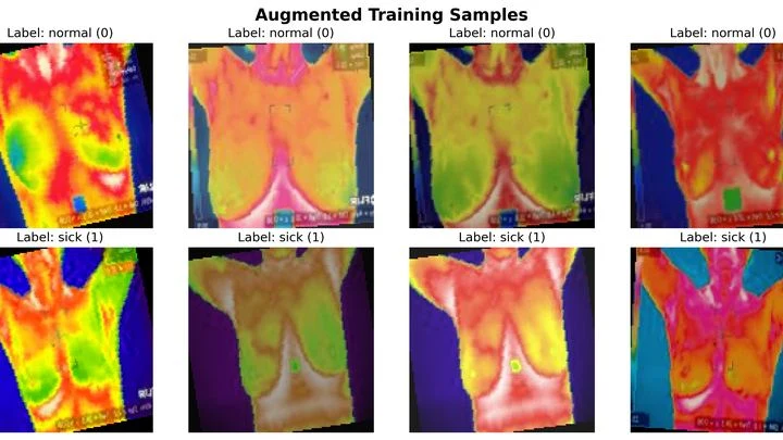
The hot air above asphalt shimmers, betraying the heated surface. Medical thermography works in a similar way: the skin over a tumor is often slightly warmer than healthy skin — by just fractions of a degree. Computer programs try to catch this difference and determine whether it's dangerous.
Two nearly identical locks are opened by the same pick. They look alike on the outside, but inside the mechanisms differ. Much the same can be said for the two main types of non-small cell lung cancer — adenocarcinoma and squamous cell carcinoma. Under a microscope, they can be confused, yet they require different treatments. That's where the quantum "magnifier" comes in.
An ordinary camera captures light reflected from objects and builds a picture from it. But if the object is hidden behind an obstacle or does not reflect light, radio waves come to the rescue: they pass through walls and fog, and a computer reconstructs the image from scattered signals.
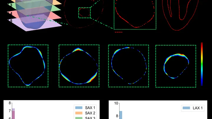
The heart is not a perfect sphere; its shape changes with every beat. To build an accurate 3D copy for a computer model, doctors typically take dozens of MRI slices. But that's like assembling a watermelon from thin slices: the fewer slices, the more guesswork.
A new approach teaches a neural network to understand the heart's shape from sparse contours.
A stroke hits suddenly, but before the strike, the brain sends quiet signals. Blood vessels stop smoothly regulating blood flow, like a thermostat that starts overheating or overcooling a room without reason. This imbalance is invisible on ordinary wearables, but it can be caught by listening to several body rhythms at once.
A doctor stares at a gray-white smudge on the screen. It's the face of an unborn child. One shadow in the right place, and you can plan surgery in time. But not everyone sees that shadow: cleft lip or palate is rare, and a young specialist might go years without ever spotting one.
An electrocardiogram is like a recording of an engine's hum: an experienced mechanic can hear when something's off. A heart ultrasound shows the engine itself from the inside, but the machine is pricey and specialists are scarce.
A particle detector at the Large Hadron Collider runs for years without stopping. Its sensors produce mountains of data every second, and among it all, you need to spot a fault quickly, like overheating or a broken channel. In the past, simple rules did this, but they often made mistakes.
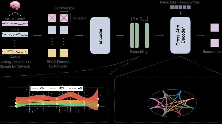
The brain isn't one big computer: it's more like an orchestra, where different groups of musicians play their parts. Neural networks for memory, attention, or emotions are constantly talking to each other. In Alzheimer's disease, this orchestra starts to go off-key: some groups fall silent, others play out of sync.
An electrocardiogram is like a long strip of paper on which the heart leaves its signature. A doctor studies it to catch rhythm disturbances, but when the recording lasts for days, the strip stretches for miles. The eye tires, attention wanders, and rare but dangerous irregularities can slip by.
AI-HEART works like a tireless assistant that scans the entire strip without a break.
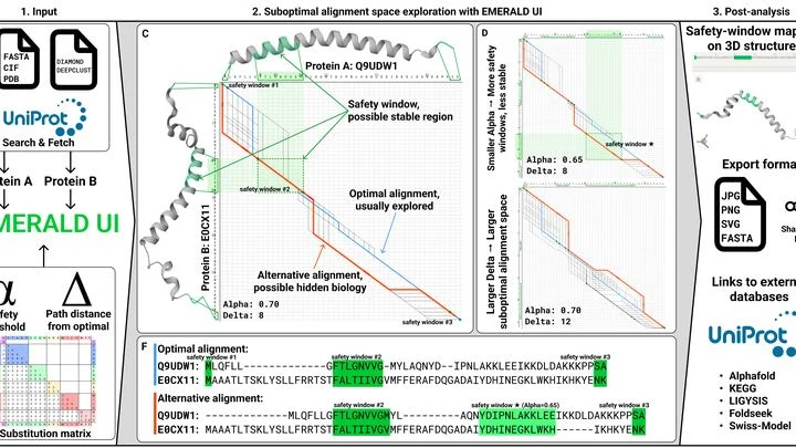
Proteins are the molecular machines of life. They fold into complex three-dimensional shapes, like a sheet of paper into origami. But if you fold the paper a bit differently, you get a different figure. Likewise, a protein can take on several forms, and that determines how it works.
Usually, scientists compare proteins by their sequences — chains of amino acids.
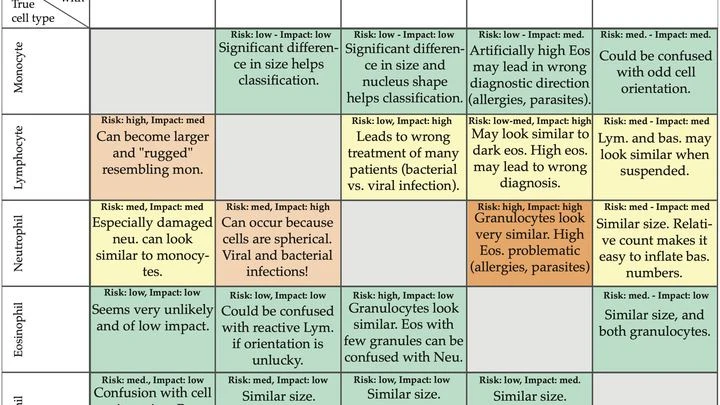
A drop of blood lands on a tiny sensor, and within a minute the doctor knows how many different defenders are in it. That's the future of blood testing, brought closer by Danish engineers.
Instead of a regular microscope with lenses, they built a lensless device. A cell flows over the sensor, which records a pattern of dark and light rings — a hologram.
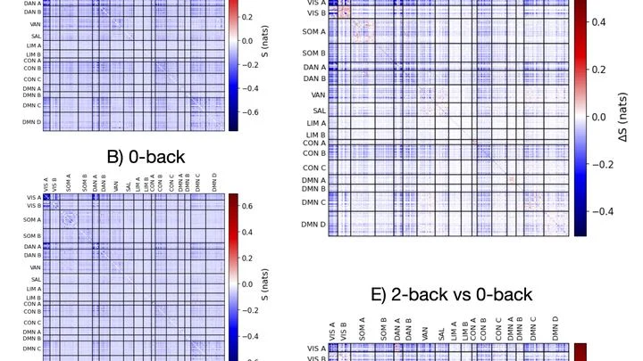
Imagine an office where employees exchange notes. When there are few tasks, everyone works independently. But when an urgent order arrives, some departments start actively messaging others, and the overall work becomes more coordinated. That's roughly how the brain behaves when we try to hold several things in mind at once.
An aneurysm is a weak spot in a blood vessel wall that bulges out like a bubble on an old bicycle inner tube. If the bubble bursts, blood floods into the brain. Doctors need to know how stressed the aneurysm wall is to decide whether to operate or just monitor.
In a dark room, on a table, sit a laser pointer, a piece of glass with tissue, and an ordinary phone camera. Light passes through the sample, and the image shows not cells but a pattern of bright and dark stripes, like the shadow of leaves on a wall.
A detective is looking for a criminal in a file of mugshots, but the portraits are mixed up. Usually, they expect a man with a gray beard, but the culprit is young and clean-shaven. Similarly, doctors often fail to recognize Alzheimer's disease in those who show unusual symptoms — not memory loss, but problems with vision or speech.