Cells around obstacles: why pillar size decides where swirls form ✛
Cells in living tissue are like fibers in a piece of cloth: they align in one direction. If they encounter obstacles, such as two pillars, they have to go around them. At places where the direction changes sharply, special points form, which scientists call topological defects. They are like swirls in a water stream when it flows around stones.
The researchers grew a layer of muscle cells on a surface with a round pit and two pillars. The pillar diameter varied from 0.1 to 0.4 mm. It turned out that at diameters of 0.3 and 0.4 mm, exactly two defects always form in the layer. Each defect has a charge of -1/2, meaning that the orientation of cells around it twists by half a turn. Two such defects together give a total charge of -1, which matches the mathematical rule for a region with two holes.
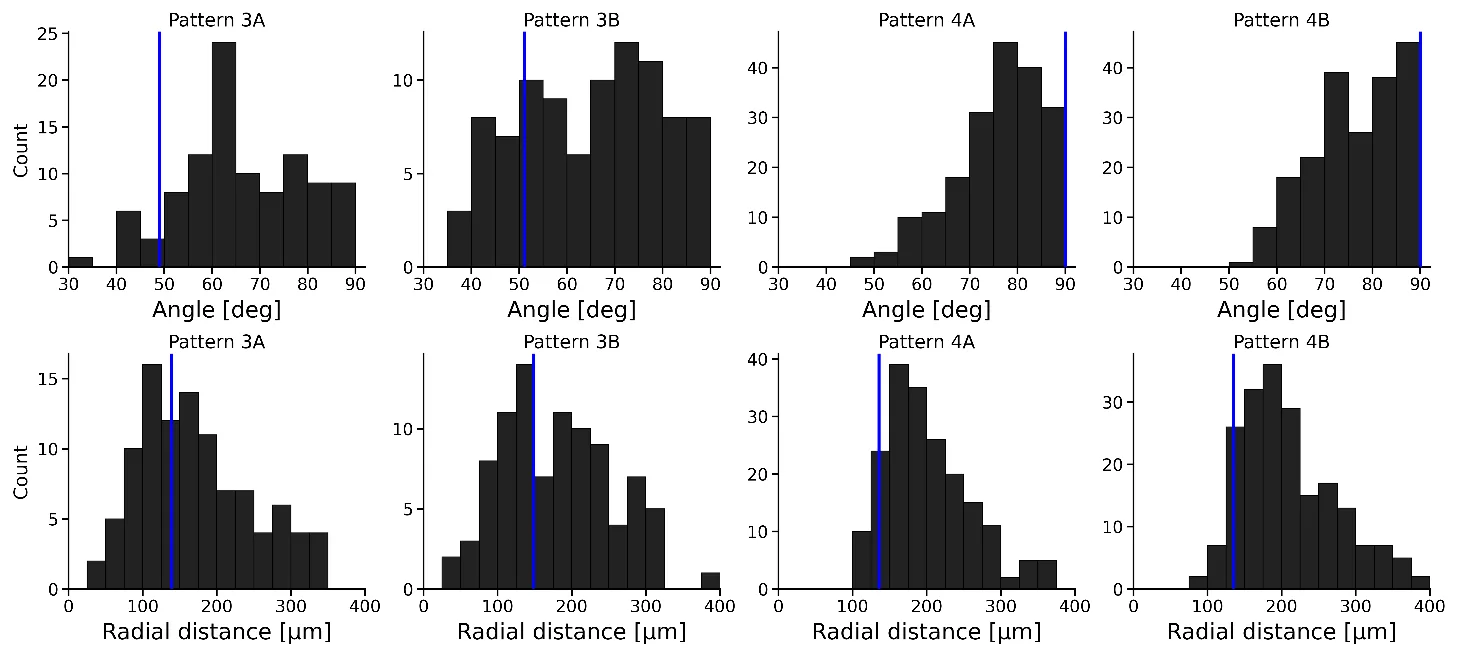
The scientists noticed that the position of the defects depends on the pillar size. With large pillars (0.4 mm), the defects lie almost on the straight line connecting the centers of the holes. With smaller ones (0.3 mm), they shift toward the center and deviate. To understand why this happens, they built a model in which cells behave like a liquid crystal. Calculations showed that for small pillars, a horizontal arrangement of defects is favorable; for medium ones, a tilted arrangement; and for large ones, a vertical arrangement. The predictions matched the experiments to within 60 μm, which is less than the size of a single cell.
This discovery helps understand how tissues behave around blood vessels, ducts, and other cavities. Knowing the size of obstacles, one can predict where defects will form, and thus how the tissue will contract and move.
In the human heart, two -1/2 defects are found between the ventricles, and their position matches predictions for a triply connected domain.
In Stanisław Lem's novel Solaris, the ocean creates complex structures that react to the geometry of the station; similarly, cell layers reorganize their orientation around obstacles, generating topological defects.