Defects in the Fabric: How Obstacle Size Dictates Symmetry ✛
Context
Topological defects in cell layers govern apoptosis, wound healing, and morphogenesis. Previous studies mostly focused on regions without holes or with a single hole, where the total charge is non-negative. However, biological tissues often surround multiple obstacles, such as blood vessels or cavities. In such regions with two or more holes, the Euler characteristic is negative, and defects with negative charge must arise. This work is the first to experimentally and theoretically investigate stable configurations of two -1/2 defects in a triply connected domain, which is important for understanding tissue mechanics around internal obstacles.
Methods
C2C12 myoblasts were cultured on polydimethylsiloxane substrates with a circular well of 1 mm diameter and two symmetric cylindrical pillars with diameters ranging from 0.1 to 0.4 mm. Cell orientation was determined from phase-contrast images using the OrientationJ plugin, and defects were identified by local minima of the order parameter. Theoretically, the orientation field was constructed analytically via a holomorphic function satisfying tangential anchoring boundary conditions. The Frank energy was minimized numerically using Newton's method, varying the positions of two -1/2 defects.
Results
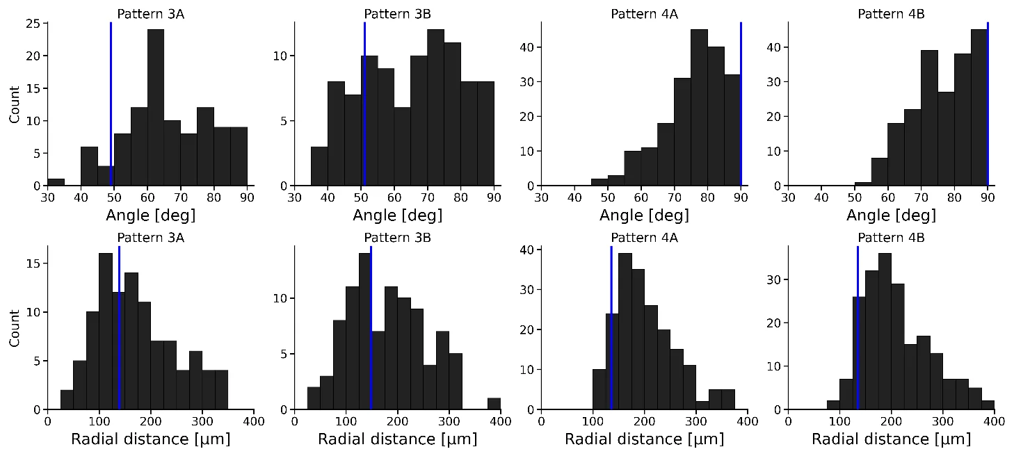
For obstacle diameters of 0.3 and 0.4 mm, most samples exhibited exactly two -1/2 defects, and the total topological charge was -1, as required by the Poincaré–Hopf theorem. For smaller obstacles, anchoring at the boundaries was disrupted, and additional defects appeared. The spatial distribution of defects showed that at a diameter of 0.4 mm, they were localized near the vertical axis, while at 0.3 mm they shifted toward the center and deviated from the axis. Theoretical analysis revealed bifurcations: for obstacle radii less than 0.18, the horizontal configuration is most stable; from 0.18 to 0.38, the off-axis configuration; and above 0.39, the vertical configuration. Experimental distribution peaks coincided with theoretical energy minima to within 60 μm.
Implications
This work shows that the size of internal obstacles governs the symmetry of cell alignment through topological constraints. Unlike previous studies in simply connected domains, where defect positions were determined by boundary curvature, here the interaction of defects with holes creates multiple critical points in the energy landscape. This explains how tissues can generate different force patterns during morphogenesis or organ movement by changing the size of cavities. The obtained analytical relationship between geometry and defect configuration provides a basis for predicting tissue mechanics with obstacles.
Future development
The theoretical model can be extended to non-horizontal anchoring and defect dynamics, incorporating stochastic initial conditions. Experiments with other cell types and obstacles of different shapes will help test the universality of the findings. Also promising is the study of three-dimensional analogs, such as disclination lines in cardiac muscle, where two -1/2 defects are observed between the ventricles.
Impact
The results are applicable in tissue engineering for designing scaffolds with desired cell orientation, as well as in understanding tumor growth around blood vessels and epithelial tissue rupture.
Next steps
Conduct experiments with obstacles of intermediate sizes to map the bifurcations in detail. Include in the model the dynamics of defect pair annihilation and variability of initial states.
Key open problems
This work links the geometry of the domain to topological defects in biological tissues, which is important for understanding morphogenesis and organ mechanics. An open question remains how cells sense obstacle curvature and how this affects their contractile activity.
The knowledge machine's view
This section is addressed to the author of the paper and their colleagues. Neighbouring works were found by meaning-based search across our archive, not by word overlap. We do not review: we show the field around the work.
What the work does
This study experimentally and theoretically investigates the configurations of two topological defects with charge -1/2 in a cell monolayer surrounding two cylindrical obstacles. It shows that the obstacle size determines the symmetry of the defect arrangement: from horizontal, through off-axis, to vertical. A theoretical model based on Frank energy reproduces the observed defect positions with an accuracy of 60 μm.
Where its strength is
The strength of this work lies in combining experiments with analytical theory for a triply connected domain, which revealed bifurcations in defect positions as the geometry changes. The established relationship between obstacle size and defect symmetry is a concrete result that can serve as a foundation for further research.
Where it could go next
- It might be interesting to compare the effect of boundary curvature on defect positions with results for simply connected domains, where curvature dictates defect size, as in the work on celestial mechanics of cells.based on: Celestial Mechanics of Cells: How Curvature Dictates Size arXiv:2607.07102
- A related avenue is using topological constraints to control cell behavior: in the work on cosmic strings in nanocomposites, similar defects are used to create structures, which might suggest how to use defect configurations to guide tissue growth.based on: Cosmic Strings in a Nanocomposite: How to See the Echo of the Big Bang arXiv:2607.14697
- It could be useful to consider defect dynamics when obstacle shape or number changes, drawing on the approach from the work on how cells build the growth boundary, which studies cell interactions with geometric constraints.based on: How Cells Build a Growth Boundary arXiv:2603.28630
Worth trying
- Test whether the predicted bifurcation persists for non-circular obstacle shapes, such as elliptical ones.
- Investigate the dynamics of transitions between configurations when obstacle size changes slowly in a living monolayer.
- Use the identified relationship to create substrates with predetermined defect arrangements and test their influence on collective cell migration.
Why this may matter
Understanding how obstacle geometry governs topological defects could provide tools for controlling cell organization in tissue engineering and for explaining morphogenetic processes where tissues surround multiple cavities.
Nearby works in our archive
- Light Twists into Vortices Inside Tumorssimilarity 0.52
- Celestial Mechanics of Cells: How Curvature Dictates Sizesimilarity 0.52
- A Family Affliction: Why Charged Lines Refuse to Respect Symmetrysimilarity 0.51
- How Cells Choose a Body Shapesimilarity 0.51
- Cell Divisions Quench Avalanches in Living Tissuesimilarity 0.51
- Cosmic Strings in a Nanocomposite: How to See the Echo of the Big Bangsimilarity 0.50
- How Cells Build a Growth Boundarysimilarity 0.49
The archive is sparse around this work: the closest review is only 0.52, against a usual 0.64. It looks like few have drilled nearby.
In the human heart, two -1/2 defects are found between the ventricles, and their position matches predictions for a triply connected domain.
In Stanisław Lem's novel Solaris, the ocean creates complex structures that react to the geometry of the station; similarly, cell layers reorganize their orientation around obstacles, generating topological defects.
Key numbers
- diameter of the outer well: 1 mm
- obstacle diameters: 0.1–0.4 mm
- agreement between theory and experiment: 60 μm
- total topological charge: -1
- number of defects: 2