How to Make Tissues Transparent for Microscopy express
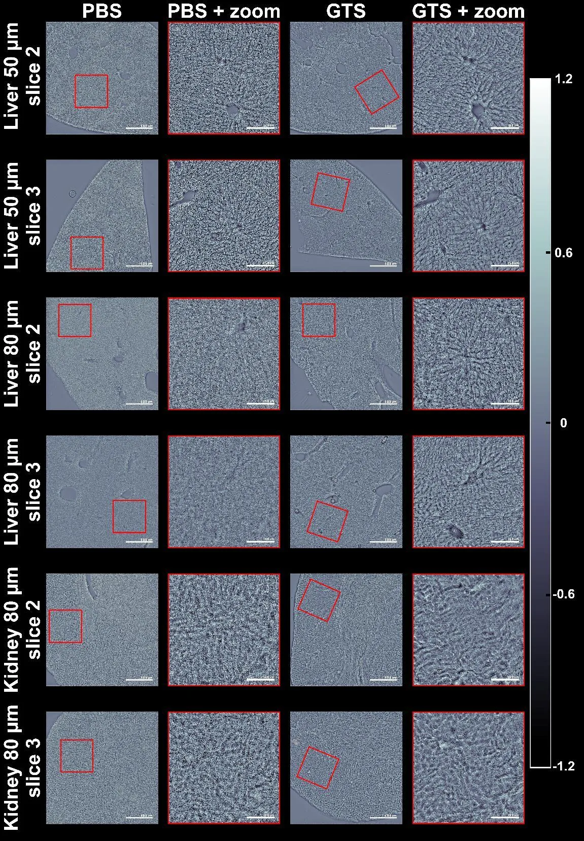
A thick slice of tissue under a microscope looks like fogged glass: light scatters and details are lost. Usually, to see cells, you stain them or cut them into very thin slices. But there's a simpler way — make the slice itself transparent, as if you've wiped the glass clean.
A solution of 60% glycerin and 10% tartrazine (a yellow food dye) immerses a piece of mouse liver or kidney in an "invisible" medium. Within minutes, the tissue becomes more transparent, and the microscope reveals its internal structure without any dyes. The researchers tested the method on sections 50–80 micrometers thick — about the thickness of a human hair.
The results were assessed not by eye but by image clarity: after treatment with the solution, the "noise" from light scattering decreased, and cell boundaries became more distinct. The solution can be stored for months at room temperature, and after analysis, it can be washed off with regular saline, allowing the same sample to be used for other studies.
The method was compared with the popular commercial preparation Ce3D — our solution proved cheaper, safer, and easier to work with. This approach could speed up routine diagnostics in labs that examine hundreds of tissue samples every day.
Original abstract
as written by the authors arXiv:2602.09732
Thick tissue sections strongly scatter and absorb light, which limits transmission-based label-free examination via quantitative phase imaging (QPI) modalities. Here we introduce a simple, room-temperature optical clearing medium - glycerol and tartrazine solution (GTS; 60 percent glycerol, 10 percent tartrazine) - that increases the transparency of 50-80 micron murine liver and kidney slices while preserving tissue morphology and enabling rapid, label-free quantitative phase imaging. Using Fourier ptychographic microscopy (FPM) and lensless digital holographic microscopy with pixel super-resolution (LDHM-PSR), we demonstrate markedly improved high-throughput visualization of microstructural features after GTS immersion compared with phosphate-buffered saline (PBS). The improvement is confirmed quantitatively by a significant increase in the one-pixel-lag autocorrelation of phase-gradient values, indicating reduced scattering-driven phase artifacts and enhanced structural continuity in reconstructed phase maps. We further show that GTS is stable for months at room temperature, provides instant yet durable clearing that improves over time, and can be removed by brief PBS washing, enabling downstream multimodal analyses on the same specimen. Finally, we benchmark GTS against the state-of-the-art clearing agent Ce3D, highlighting GTS as a low-cost, safe, operationally straightforward alternative with strong potential for routine, high-throughput QPI-based histopathology workflows.
Tartrazine, used in the solution, is the same dye that gives some popular soft drinks and snacks their bright yellow color.