Contrast in imaging is the difference in signal intensity between adjacent regions of an image — the reason a tumor shows up as a dark or bright blob against healthy tissue. Without contrast, a medical scan is a flat gray wash; with it, boundaries and textures become visible. The concept is central to radiology, microscopy, and astronomy: any technique that boosts contrast makes faint or subtle features detectable.
History
The term entered medical imaging with the advent of X-rays in the 1890s, but the systematic use of contrast agents began in the 1920s with iodine compounds for angiography.
How it works
Contrast arises from differences in how materials absorb, scatter, or emit radiation. In X-ray imaging, dense tissue (like bone or a calcified tumor) absorbs more photons, casting a shadow; in MRI, contrast agents alter the magnetic relaxation of nearby water molecules, brightening or darkening specific areas. The image is a map of these intensity differences, and the eye or a computer algorithm reads them as structure.
In practice
Contrast agents are injected into patients to highlight tumors, blood vessels, or inflammation in CT, MRI, and ultrasound scans; in microscopy, fluorescent dyes provide contrast to reveal cellular structures.
Fun fact
In some imaging techniques, contrast can be enhanced by subtracting a pre-contrast image from a post-contrast one, leaving only the agent's signal — a trick that makes even tiny lesions stand out.
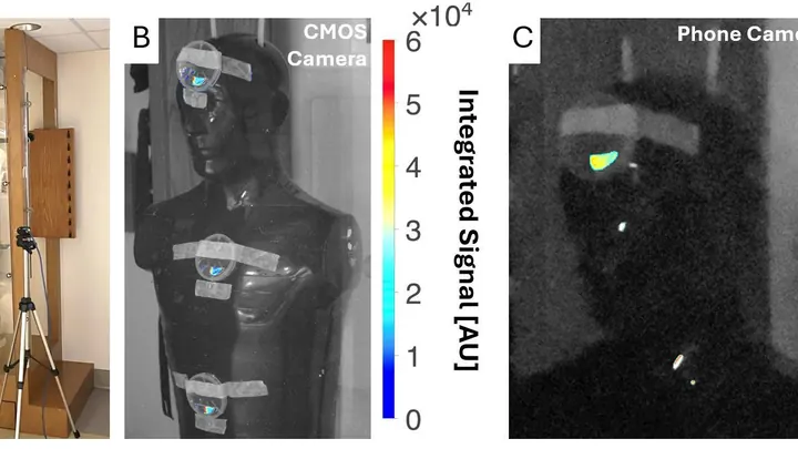
A crystal the size of a matchbox was placed under the beam of a clinical accelerator. Half of its surface was covered with a pattern of nanoscale bumps — tiny, smaller than the wavelength of light. The other half was left smooth. The difference was striking: the coated part produced a signal 4.1 times brighter.
A surgeon removes a breast tumor, but how do you know no cancer cells are left at the edge? Right now, the tissue goes to a lab, and the answer comes back days later. If the margin is not clean, a second surgery is needed. The new method promises to give the answer right in the operating room, while the patient is still on the table.
A drummer stays silent, but from the pauses you can tell where he didn’t hit. Similarly, physicists built an image by catching not light, but moments of absolute darkness. The picture emerged from silence.
In the experiment, they shone light from an ordinary lamp onto the object.
In the operating room, the surgeon illuminates the tumor with a special agent that makes it glow. But how do you know whether it glows brightly because there are many cancer cells or simply because it lies close to the surface?
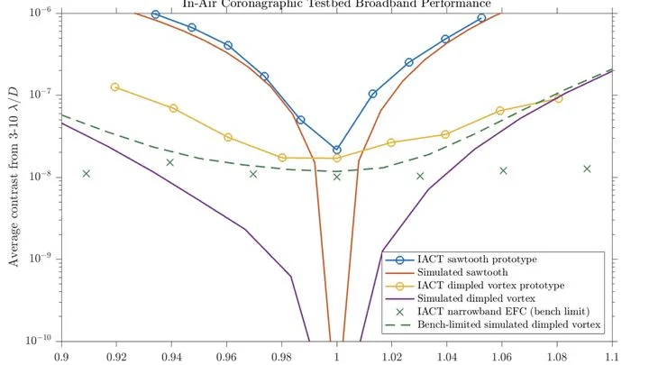
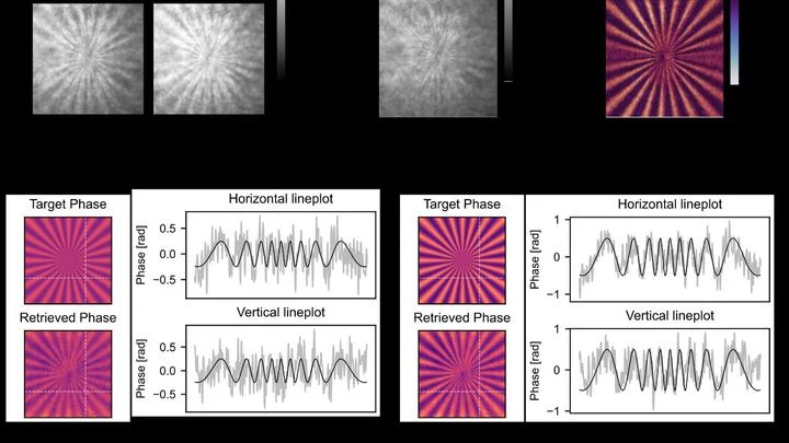
A star shines billions of times brighter than its planet. To see the planet, you need to quench the starlight, like shading your eyes with your hand to spot an airplane against the sun. Telescopes use a mask with a clever pattern that twists the star's light into a ring and cancels it out, while the planet's light passes through almost untouched.
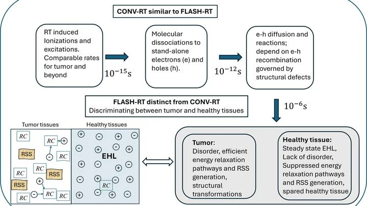
Two cities: one with neat, straight streets, the other with a tangle of alleys. If you send a powerful flood of water through both, in the first the water quickly drains away through channels, while in the second it swamps everything. That's roughly how tissues behave under ultrafast radiation therapy, known as FLASH.
Gas is nearly invisible, but it has a temperature, and you can read it from how the gas 'shadows' the thermal background. In the experiment, a camera looks at a warm screen through a jet of carbon dioxide. If the gas is cooler than the background, it appears as a dark spot; if warmer, it appears bright.
A glass marble in a glass of water is nearly invisible, but if you look through it at a checkered tablecloth, the lines behind it appear distorted. Transparent cells behave the same way: light passes through them but is slightly slowed and changes direction.
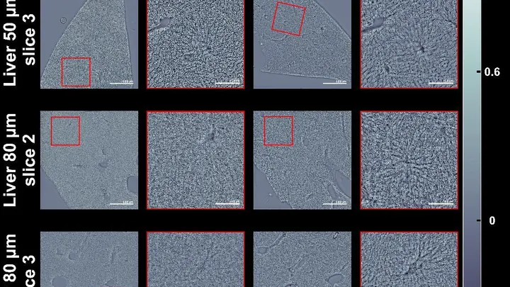
A thick slice of tissue under a microscope looks like fogged glass: light scatters and details are lost. Usually, to see cells, you stain them or cut them into very thin slices. But there's a simpler way — make the slice itself transparent, as if you've wiped the glass clean.
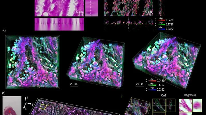
Light passing through a stained cell loses some of its strength, like a flashlight beam cutting through a glass of tea. The denser the color, the more light is absorbed. But until now, a microscope could only show a flat shadow of this process, not its volume.