Staining is a laboratory technique used to add color or fluorescence to biological samples, enhancing contrast so that structures become visible under a microscope. Since most cells are nearly transparent, staining allows researchers to distinguish nuclei, organelles, proteins, or even specific DNA sequences. It is essential in histology, pathology, and cell biology for diagnosing diseases and studying tissue architecture.
History
The technique dates back to the 19th century; the first synthetic dyes were used in microscopy in the 1850s, and the Gram stain, still used today, was developed by Hans Christian Gram in 1884.
How it works
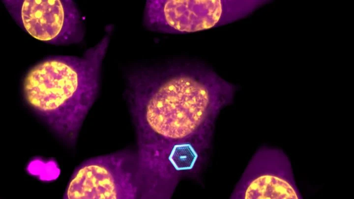
Stains work by binding selectively to cellular components. For example, hematoxylin binds to nucleic acids (blue), while eosin binds to proteins (pink). Fluorescent stains, like DAPI or GFP, absorb light at one wavelength and emit at another, enabling multi-color imaging. The process typically involves fixing the sample, applying the stain, and washing away excess, followed by microscopy.
Some stains, like the Golgi stain, reveal only a small percentage of neurons in a sample, which paradoxically makes individual cells easier to study against a clear background.
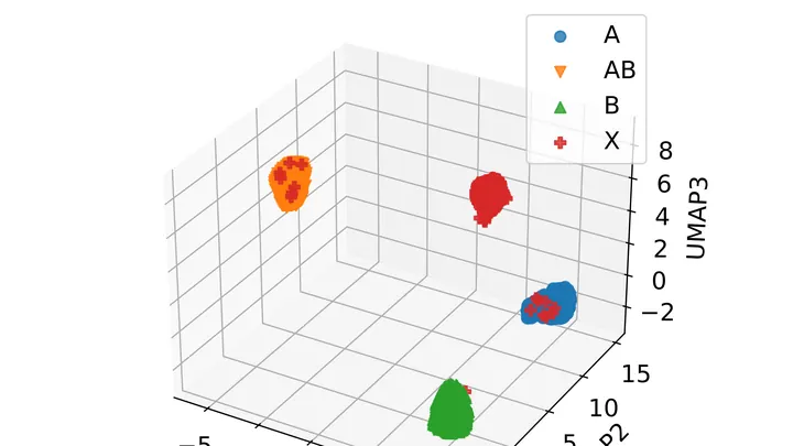
A stain is found at a crime scene. The expert takes a swab, and a few hours later the instrument outputs a list of proteins that made up the fluid. Previously, a rapid test could be wrong if multiple fluids were present: blood mixed with saliva, and the strip gave a false result.
A thin section of eucalyptus lies under the microscope objective, and white light, passing through two polarizing filters, turns the wood into a mosaic of purple, yellow, and blue patches. These colors are not random: like letters on a map, they show the direction of the invisible cellulose threads inside the cell walls.
Studying distant exoplanets, scientists look for signs of life — for instance, water and carbon — and signs of technology: radio signals, megastructures. But between these clues there is a yawning gap. If intelligence hasn't created machines or they've vanished, we won't notice it.
Biologists often tag cells with glowing molecules — it's like searching for a dim light in the smoke. The light quickly fades, and the smoke creates interference.
A pathologist stains a tissue section with hematoxylin and eosin — cells turn blue and pink, and under the microscope you can see their shape and arrangement. But which genes are active in each cell cannot be read from such an image. That requires a separate, expensive assay that reveals the activity of thousands of genes at once, but it is rarely performed.
A mole on your skin looks like a mosaic of colored pieces. Each piece is a small patch of the image where color and texture barely change. Such pieces are called superpixels. If you connect them with lines, you get a network that looks like a subway map: stations are the pieces, and lines are the connections between them. The weight of each line shows how strongly two pieces are related.
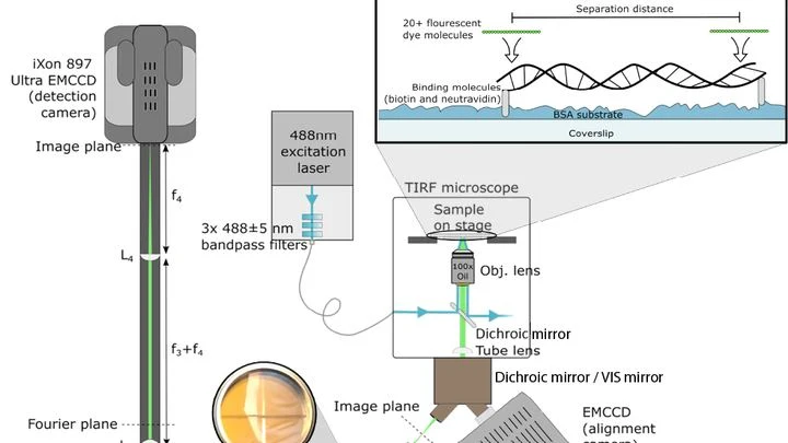
A conventional light microscope hits a fundamental limit: it cannot distinguish two points closer than 200 nanometers — roughly the thickness of a soap bubble divided by a hundred. To see deeper, scientists have made molecules in the sample blink or fade, but these tricks are slow and harmful to living cells.
A sheet of graphene is a mesh of carbon atoms one atom thick. Even the slightest contamination shows up on it, like dust specks on eyeglass lenses. Ten years ago, researchers learned to hide graphene between layers of boron nitride, like placing a fragile sheet between two pieces of plastic wrap.
A scanning tunneling microscope probes a molecule with an ultra-fine tip and maps its relief. But the image is flat, while the molecule is three-dimensional. How do you guess a person's pose from their shadow on a wall?
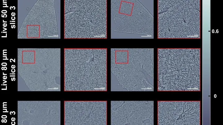
A thick slice of tissue under a microscope looks like fogged glass: light scatters and details are lost. Usually, to see cells, you stain them or cut them into very thin slices. But there's a simpler way — make the slice itself transparent, as if you've wiped the glass clean.
In winter, puddles freeze, and ice cracks the asphalt. Inside a living cell, freezing does something similar: ice crystals tear its membrane. To prevent this, the cell is soaked in a special solution, like antifreeze for a car. But precision is key: too little solution and ice still forms; too much and the solution itself poisons the cell.
In a dark room, on a table, sit a laser pointer, a piece of glass with tissue, and an ordinary phone camera. Light passes through the sample, and the image shows not cells but a pattern of bright and dark stripes, like the shadow of leaves on a wall.
A pathologist looks through the eyepiece and sees a pink-purple tissue section — the classic hematoxylin and eosin stain. The dyes bind to nuclei and cytoplasm, and the color tells the doctor about the disease. But preparing the slide itself takes hours: fixation, paraffin embedding, sectioning, staining.
This new device skips the dye.
You can trap light in a cage smaller than a speck of dust. Photonic crystals are lattices that stop light from scattering and force it to run in circles. Previously, such structures were grown on a bulky substrate, making them too large for living cells.
Every cell is a miniature tuning fork. A sharp magnetic pulse makes it 'ring', and the decay of that sound depends on what's inside: viscosity, density, composition. A tiny diamond with a structural defect acts like a hypersensitive microphone, picking up magnetic echoes from individual cells.
Many molecules, like amino acids, are like musical notes: their left and right versions sound almost identical, yet the difference is critical. One heals, the other is poison. Picking up this difference is hindered by inescapable noise, like in a room where a fan is loudly running. Regular spectroscopy misses the nuances.
Inside every living cell, chemical transformations are constantly taking place. Some of them create unstable particles whose electrons behave like paired magnets. Their interaction resembles a dance: each movement of the pair responds to external magnetic fields, as if to a changing rhythm.