How a Light Beam Is Tailored to Brain Depth express
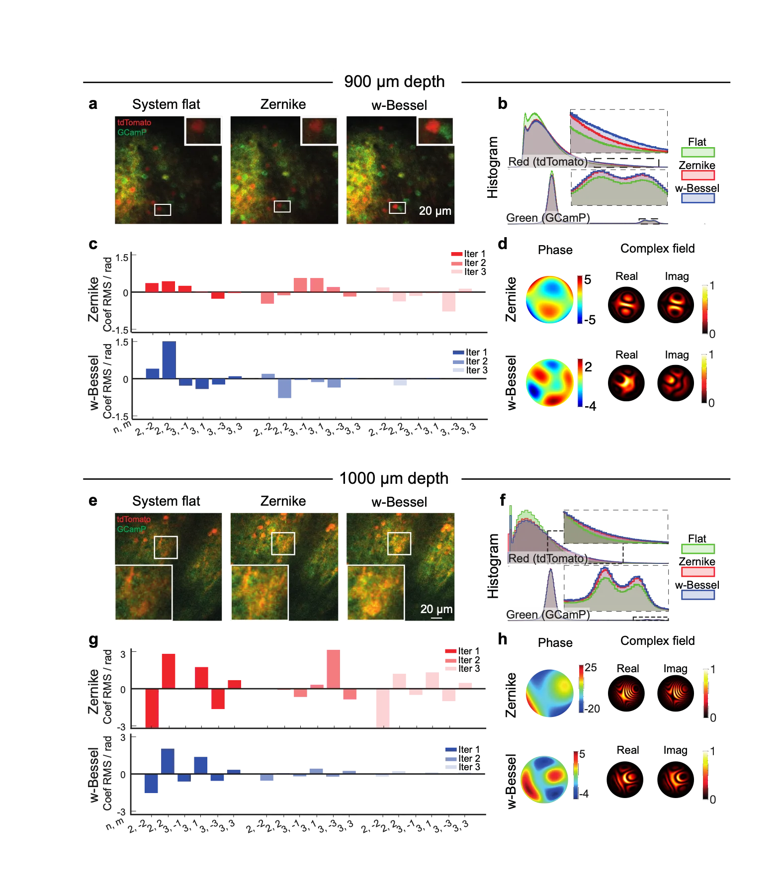
The laser beam in a three-photon microscope works like a flashlight shining through murky water. The deeper you need to illuminate, the more the water scatters light and the dimmer the picture gets. To avoid overheating the tissue, laser power is limited, so you have to squeeze every bit of performance out of the beam shape and the aberration correction method.
Usually, the beam size is fixed in advance, and aberrations are corrected using a set of standard "lenses"—the so-called Zernike modes. But, as the authors found, when the beam is reduced, these modes stop being independent: correcting one starts messing up another. Alignment drags on, the focal spot shifts, and the image suffers.
The solution is to dynamically pick both the beam size and the set of correction modes for each depth. The beam is made narrower, and the modes are specifically tailored to that narrow beam. Then alignment converges faster, the focus stays put, and the signal from deep brain layers gets stronger. In live mice, this yielded sharper images of nerve cells at great depths while keeping laser power safe.
Three-photon microscopy uses infrared light, which scatters less in biological tissues than visible light, so it can peer into the brain at depths beyond a millimeter.