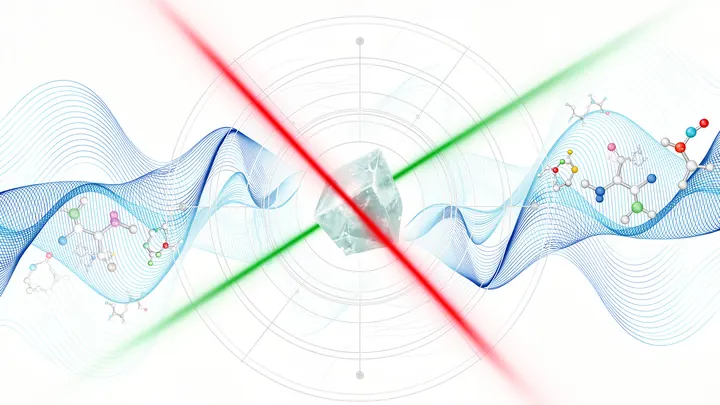
An optical microscope forms an image by refracting visible light through a series of lenses. Resolution is limited by diffraction: according to the Rayleigh criterion, two points are distinguishable if the distance between them is at least half the wavelength divided by the numerical aperture of the objective. Modern methods—fluorescence microscopy, confocal, super-resolution (STED, PALM)—overcome this limit, allowing visualization of structures down to single molecules.
History
Zacharias Janssen is credited with constructing a compound microscope (1590). Antonie van Leeuwenhoek (1674) achieved 300x magnification with single lenses, discovering microorganisms. Robert Hooke in 'Micrographia' (1665) described cells. In 1873, Ernst Abbe developed the theory of image formation and the sine condition. Carl Zeiss and Otto Schott established industrial production of high-quality objectives. In the 20th century, Frits Zernike invented the phase-contrast microscope (1935, Nobel 1953), Marvin Minsky patented the confocal principle (1957). Super-resolution emerged in the 2000s: STED by Stefan Hell (2014 Nobel), PALM by Eric Betzig and Harold Hess.
How it works
A beam of light from the source is focused by the condenser onto the specimen. Scattered and directly transmitted light is collected by the objective, which forms a primary image in the plane of the field diaphragm. The eyepiece further magnifies it for viewing by eye or transmission to a detector. Contrast in brightfield microscopes arises from absorption or scattering; phase contrast converts a phase shift into an amplitude change. In fluorescence microscopes, the specimen is illuminated at the excitation wavelength, and emitted radiation is separated by a dichroic mirror and filters. In confocal microscopy, a point source and a detector with a pinhole provide optical sectioning. Super-resolution overcomes the diffraction limit by turning off fluorophores at the periphery of the excitation spot (STED) or by precise localization of single photoactivatable molecules (PALM/STORM).
In practice
Used in biology and medicine for cell analysis, in materials science for inspecting microchips, and in forensics for examining fibers and prints.
Fun fact
With an optical microscope, you can not only look but also move objects: laser tweezers can hold and move individual cells or bacteria.
A stroboscope in a dark room picks out fleeting shadows from the crowd, shadows the eye cannot catch. A primordial black hole the size of a virus, passing in front of a distant star, briefly magnifies its light for a few seconds, like a magnifying glass held over a bulb.
A thin section of eucalyptus lies under the microscope objective, and white light, passing through two polarizing filters, turns the wood into a mosaic of purple, yellow, and blue patches. These colors are not random: like letters on a map, they show the direction of the invisible cellulose threads inside the cell walls.
The electron beam of a microscope usually flies through emptiness, like a courier on an empty highway. Any air molecule is a bump that makes it bounce and lose direction. So before, to examine a sample, you had to freeze it, dry it, or coat it with metal—and still you could only look in a vacuum.
A doctor looks at a screen and sees two images of the same part of the body. One shows where blood is rich in oxygen, the other shows how fast it rushes through the finest vessels. Previously, these images were made by different devices, and combining them was difficult. Now engineers have built a device that takes both images simultaneously.
Light passing by a tiny structure changes direction. If the structure is smaller than the wavelength of the light, it acts like a lens and focuses the light into a spot smaller than the wave itself.
A doctor looks at a screen and sees two images of the same part of the body. One shows where blood is rich in oxygen, the other shows how fast it rushes through the finest vessels. Previously, these images were made by different devices, and combining them was difficult. Now engineers have built a device that takes both images simultaneously.
Light passing by a tiny structure changes direction. If the structure is smaller than the wavelength of the light, it acts like a lens and focuses the light into a spot smaller than the wave itself.
Inside a microscope that sees individual atoms, it's cold and silent: the temperature is around -268°C, and the interior is in perfect vacuum. To place individual iron atoms on such a stage, you usually need a direct 'corridor' from the evaporator to the sample. But in the coldest instruments, that corridor is blocked by thermal shields, making deposition a puzzle.
Light in an optical fiber is like a stream: it flows inside a glass thread and doesn't spill out. A thick thread carries many streams at once, a thin one only one. Astronomers often need to pour light from a thick fiber into several thin ones to split it into colors.
Data zips through optical fibers at the speed of light, but the path isn't perfect: the signal gets muddled, like a voice echoing down a long hallway. The faster the speed and the longer the distance, the worse the distortion. Usually, electronics fix this, but they can't keep up with light.
An ordinary light bulb is either on or off. But tiny glowing particles, nanocrystals with rare-earth ions, have so far emitted steady light, like small flashlights. Inside such a crystal, there are thousands of emitting ions, and even if one 'gets tired,' the others keep shining. Therefore, blinking, sharp on-and-off switching, was considered impossible for them.
The eye's vascular network is not just a peripheral part of the circulatory system; it is a 'mirror' of the brain's hemodynamics, anatomically and functionally connected to it.
In a glass of murky liquid where starch is settling, the eye sees only a gray haze. Particles inside are moving, but what exactly is happening remains a mystery. Conventional optical instruments cannot penetrate such a suspension: light scatters already in the top layer.
In a glass of murky liquid where starch is settling, the eye sees only a gray haze. Particles inside are moving, but what exactly is happening remains a mystery. Conventional optical instruments cannot penetrate such a suspension: light scatters already in the top layer.
Light passing through a glass of murky water dims — some of it is absorbed. That's how many liquid-analysis instruments work: they measure how much light was lost and infer the composition from that. But if the liquid is heterogeneous, like soup with chunks, a single number isn't enough: you need to know where exactly more absorption happened.
Light passing through a glass of murky water dims — some of it is absorbed. That's how many liquid-analysis instruments work: they measure how much light was lost and infer the composition from that. But if the liquid is heterogeneous, like soup with chunks, a single number isn't enough: you need to know where exactly more absorption happened.
A synchrotron beam is like a jet of water from a hose: wide and awkward for fine work. To examine a tiny sample, you need a fine needle, not a hose. In the new setup, the needle is a glass capillary — a tube through which light glides, reflecting off the walls like a voice echoing down a long corridor.
A synchrotron beam is like a jet of water from a hose: wide and awkward for fine work. To examine a tiny sample, you need a fine needle, not a hose. In the new setup, the needle is a glass capillary — a tube through which light glides, reflecting off the walls like a voice echoing down a long corridor.
Gravitational-wave detectors are like extremely sensitive ears: they pick up the trembling of space when black holes or neutron stars collide somewhere in the universe. But even the most sophisticated "ears" cannot hear everything. The new LIGO A# project is like replacing the eardrum with a thinner, more sensitive membrane.
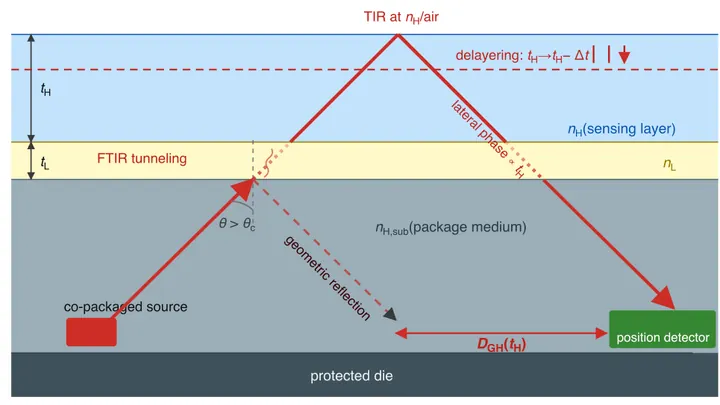
A microchip is like a layered cake: protective layers on top, secrets inside. An attacker can peel off layers one by one to get to the secrets. A new sensor monitors this with a light beam.
The beam hits the chip surface at an angle. Part of the light reflects, but not immediately: it slightly 'dives' into the top layer, like a pebble skipping across water.
Starlight passing through the atmosphere and telescope optics shimmers like sunlight dancing on water. Hidden in that ripple are planets — faint sparks next to a blinding glare. To see them, you have to freeze the ripple: make the light as smooth as glass.
A passing particle leaves a flash of light in a long transparent bar — a scintillator. Inside the bar, like threads in a rope, thin fibers are stretched to collect this light and channel it to a sensor. Particle detectors have worked this way for decades.
A camera looks at two distant sparks. They're so close together that they merge into a single fuzzy blob. It seems impossible to tell them apart — the classic limit of optical microscopy has been reached.
But quantum mechanics hints that the light field still holds a secret about where each spark sits.
The orchestra plays, but the conductor hears only a general hum. Each instrument plays its own part, but you can't tell where the flute is and where the double bass is. You need a spectrum analyzer — a device that breaks down sound by frequency.
Radiation therapy is a precision strike on a tumor, but every shot must be calibrated to within a millimeter and a fraction of a second. The problem is that existing dosimeters are either bulky, or they distort the beam themselves, or they require pauses for readout.
An array of radio telescopes is like a camera missing several lenses. Ever since Karl Jansky picked up the radio hiss of the Milky Way, astronomers have been trying to piece together a whole picture from fragmentary signals. But the antennas are spaced apart, and the mathematically “developed” image — the dirty map — ends up smeared.
A laser beam can do more than heat things up: it can also calm motion. When light enters a mirror trap, it exerts pressure on the mirrors, and if you tune the beam's color just right, that pressure becomes a gentle braking force. This is how tiny mechanical vibrations are cooled to nearly absolute zero.
An ordinary flashlight emits light whose waves oscillate in all directions at once. But pass that light through a special filter, and you get a beam that oscillates strictly in one plane: that's polarized light. When such a beam travels through biological tissue, the tissue's internal structure slightly alters the orientation of the oscillations.
A night river is dotted with tiny sparks. This isn't plankton: it's functional pollen-sensors, millimeter-sized devices that swim on their own and measure the water's chemistry.
Each particle is a tiny lab. Inside, two reagents meet: one triggers a glow upon contact with a pollutant, and the second, a colored filter, blocks out interference.
Two tiny weights, each about the mass of a grain of sand, hang in the dark at a temperature colder than deep space. They don't touch, but an invisible thread connects them — gravity. The experiment physicists are now building aims to catch the moment when that thread tugs one weight due to the motion of the other.
Organic light-emitting materials are like a busy intersection without traffic lights. Molecules receive energy but, instead of honestly giving it back as light, they often turn into dead ends: they form dark pairs (exciplexes — short-lived complexes that do not emit) or collide with each other, losing charge.
A protein in a cell is not a frozen sculpture but a restless mechanism. Its parts are constantly vibrating, and these movements determine how it grabs molecules, transmits signals, or builds the cellular scaffold. But filming the life of a single protein was hampered by two nuisances at once. First, the movements last millionths of a second — ordinary cameras simply cannot catch them.
A single-pixel camera works like an artist creating a picture through a stencil with many holes. In each frame, only a portion of the light, modulated by a mask, a spatial light modulator, reaches the single sensor, and then the computer reconstructs the full image by assembling this mosaic with computational algorithms.
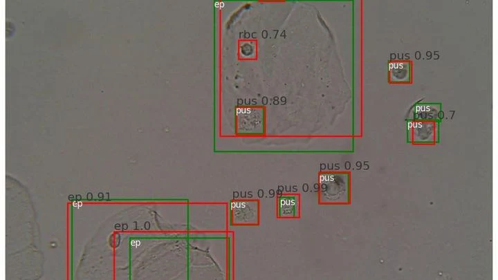
A drop of urine under a microscope isn't a flat puddle but a multi-layered volume. Like a stack of transparent pages: if you focus on one, the neighboring ones blur. The microscope's light only shows one depth layer sharply. To see all the cells, a lab technician had to take a series of snapshots, manually adjusting focus — tedious and slow.
Fiber optics transmit data with light of different colors. These beams race at nearly the speed of light. Normally, all signals go the same way, creating a jam and interference, like an orchestra where musicians are trying to outshout each other.
Imagine photographing a statue in dim light. You face a dilemma: take one bright shot or a series of dim frames from different angles. In the first case, quality is limited by photon noise, the graininess that arises from a lack of photons, while in the second, details overlap, making it harder to reconstruct the 3D structure.
Building a light chip is like a game where you have to guide a beam through a maze of tiny mirrors and beam splitters. Each element acts like a railroad switch: it either lets light pass straight through or directs it to an adjacent track. The goal is to produce a specific light pattern at the output while using as few 'switches' as possible.
Usually, engineers spend years finding the shortest route for each task.
Spectroscopy has always been a way to hear the unheard. Back in the 19th century, Johann Balmer deciphered the elegant lines of hydrogen — a simple numerical harmony that foreshadowed quantum theory.
Spectroscopy has always been a way to hear the unheard. Back in the 19th century, Johann Balmer deciphered the elegant lines of hydrogen — a simple numerical harmony that foreshadowed quantum theory.
Dozens of copper coils in an MRI scanner pick up the faint response from tissues, like microphones trying to catch a whisper in a noisy hall. Each microphone drags a thick bundle of wires behind it. The wires press on each other, create electrical interference, heat up, and add risk for the patient.