How a neural network catches a chemical mark on a single molecule express
A chef tastes a sauce and knows: it needs a pinch of salt. But toss that same pinch into a lake, and no one will notice the difference. The same goes for proteins: adding a single hydroxyl group (oxygen plus hydrogen) changes how a molecule works, yet spotting that change among thousands of other atoms is nearly impossible.
Scientists built a trap for individual molecules — a pore about the size of a virus, into which an electric field pulls peptide fragments. At that moment, a laser hits them, and the scattered light shimmers differently depending on whether the peptide carries that chemical mark (hydroxylation). The difference is subtle, like two shades of gray on a cloudy day. But a neural network comes to the rescue: it scans thousands of such light patterns and learns the recurring features. After training, the program distinguishes a modified fragment from an ordinary one with up to 90% accuracy.
This approach is like reading Braille: fingers glide over barely raised dots, but the brain assembles them into letters. Here, the laser plays the fingers, a convolutional neural network plays the brain, and fleeting spectral changes play the raised dots. The technology promises to recognize ultra-weak chemical signals in single molecules, which is crucial for early disease detection where such signals are the only clue.
Original abstract
as written by the authors arXiv:2605.21084
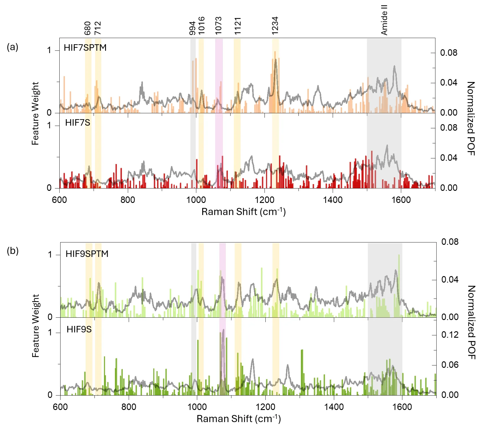
Post-translational modifications (PTMs) play essential roles in regulating protein structure, function, and cellular signalling. However, peptide level discrimination of hydroxylation at the single-molecule level remains difficult. Here, we report a particle-in-pore single-molecule surface-enhanced Raman spectroscopy (SERS) platform combined with peak occurrence frequency (POF) analysis and a one-dimensional convolutional neural network (1D-CNN) for discriminating hydroxylated and non-hydroxylated HIF peptide fragments. Three peptide pairs containing the Pro-564 hydroxylation site, with lengths of 7, 9, and 15 amino acids (AAs), were investigated. POF analysis revealed reproducible hydroxylation-dependent spectral changes in the 7AA and 9AA peptide pairs, which were attributed to changes in adsorption conformation and surface interactions. CNN-based classification achieved post-evaluation accuracies of 72.98%, 78.55%, and 89.74% for the 7AA, 9AA, and 15AA peptide pairs, respectively, with AUC values above 0.80 for all the pairs, indicating a reliable discrimination. Gradient-weighted feature visualization further showed that CNN-sensitive regions overlapped with recurrent POF features, supporting the chemical relevance of the learned classification patterns. Notably, for the 15AA peptide pair, the enhanced citrate-associated band suggests that hydroxylation can substantially alter peptide-gold nanoparticle adsorption behaviour. This adsorption-mediated effect may amplify hydroxylation-induced spectral differences and contribute to the improved discrimination accuracy despite the increased structural complexity. These results demonstrate that the particle-in-pore sensor, assisted by deep learning, can capture hydroxylation-induced spectral and adsorption changes in peptide fragments, providing a promising strategy for ultrasensitive analysis of weak PTM signatures in peptides.
Hydroxylation is the very mark that helps cells sense oxygen shortage. When oxygen is low, the HIF protein stops being degraded and triggers the growth of new blood vessels. Tumors often exploit this trick.
In Philip K. Dick's 'Minority Report,' criminals were caught by snippets of thought. Here, snippets of proteins reveal chemical marks — a step toward diagnostics where a molecule tells you about a disease before symptoms appear.