Ink Blots at the Cell Boundary: How Proteins Decide Where to Gather and Which Way to Look ✛
Two ink reservoirs stand on opposite sides of the cell junction. From them, two types of monomers flow onto the membrane, and when they meet, they form viscous droplets of complexes. The droplets barely move, but the free monomers dart around as fast as perfume molecules in a room. So begins the story of how a cell draws puncta on its boundary: discrete protein clusters that will later set the direction of tissue growth.
The model the authors built is simple in its ingredients: two types of monomers (A and B) diffuse quickly, reversibly bind into complexes of two orientations (C and C†), and those crawl slowly. Feedback enters only through reaction rates: clustering feedback K(c) enhances binding where complexes are already abundant, while cross-modulation V(c†) changes complex decay depending on the concentration of the opposite orientation. No external signals, only local rules. And here is the key: if monomers diffuse faster than complexes and feedback is strong enough, the homogeneous state loses stability. But not through a finite-wavelength Turing bifurcation, as one might expect, but through a long-wavelength mode: a slow redistribution of total mass.
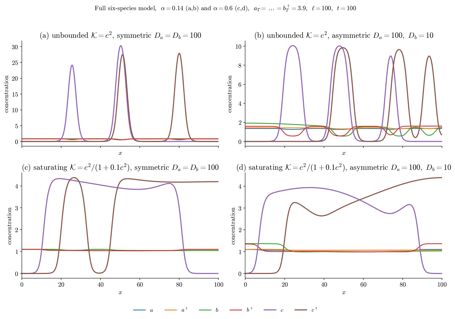
Next, the shape of the feedback decides what emerges: wide plateaus or narrow peaks. For unbounded feedback (c²), narrow peaks arise with amplitude inversely proportional to the product of the average monomer concentrations and a universal full width at half maximum of 4 arccosh(√2) ≈ 3.53 in units of the complex diffusion length. For saturating feedback (c²/(1+ρc²)), wide plateaus form, and the position of their fronts is set by the Maxwell construction, the same rule that describes phase equilibrium in physics. But the most unexpected result concerns the number of puncta. Any array of identically oriented puncta is unstable and coalesces into one big one. The coalescence speed for two peaks is proportional to the cube of the average monomer concentration and the diffusion coefficient, and when monomer diffusivities differ, it is set by the harmonic mean of their diffusion coefficients. Therefore, the number of puncta we see at a cell contact is not set by a fixed wavelength but is determined kinetically, like the slow merging of droplets on glass.
Now introduce cross-modulation: let complexes of one orientation accelerate the decay of complexes of the opposite orientation. Then clustering turns into sorting. Co-localized puncta of opposite orientations start to move apart at a speed proportional to the slope of the modulation and the amplitude of the punctum. The sorting criterion depends only on the sign of the cross-modulation, not on the shape of the clustering feedback. This means that controlling the number of puncta and their orientational segregation are mathematically separable ingredients. The cell can tune them independently, like an artist who decides separately how many spots to put on the canvas and separately which ones will look left and which right.
Thus, a model that started with two ink reservoirs gives a simple answer to a complex question: mass conservation and clustering feedback are sufficient for puncta formation, their number is set kinetically due to slow coalescence, and orientation sorting requires sign-definite cross-modulation of turnover. Today, this can be used to interpret experiments on imaging protein clusters at cell contacts: knowing monomer mobility, the model predicts how many puncta will form and what size they will be. And tomorrow, perhaps, the same ink will help draw a map of how molecular self-organization translates into tissue-level polarity.
The width of a single protein punctum in the model turns out to be universal: it does not depend on the amount of protein or the strength of feedback, but only on complex diffusivity and decay rate. This is similar to how the width of a traffic jam does not depend on the number of cars, but only on the reaction speed of drivers.