Counting Blood Cells Without Lenses express
A drop of blood lands on a tiny sensor, and within a minute the doctor knows how many different defenders are in it. That's the future of blood testing, brought closer by Danish engineers.
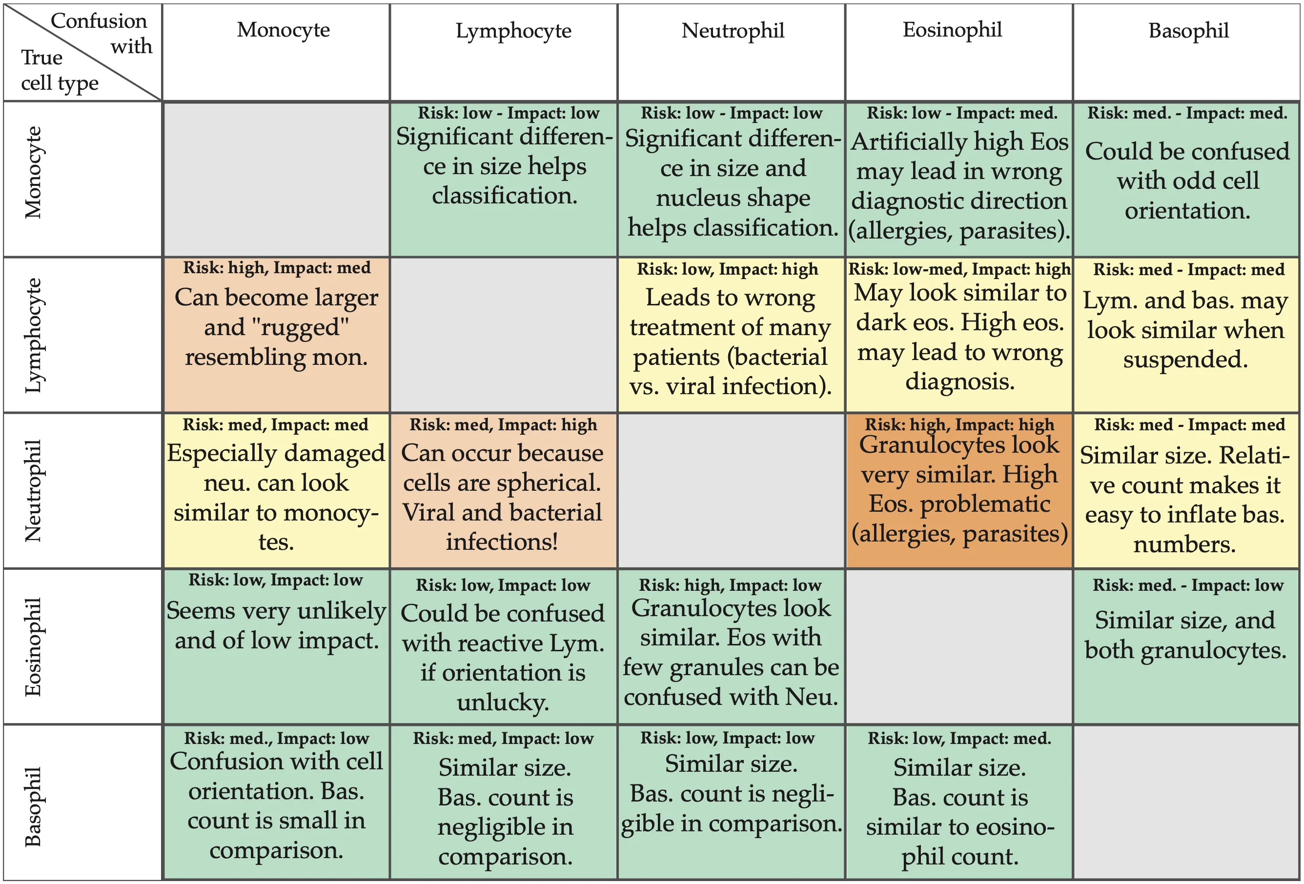
Instead of a regular microscope with lenses, they built a lensless device. A cell flows over the sensor, which records a pattern of dark and light rings — a hologram. From these rings, a neural network identifies what kind of cell it is. The first version of the device distinguished three main types of white blood cells with 89.6% accuracy. Then they simplified the design, removed the lenses, and accuracy actually improved: 92.65% for three types and 89.44% for five.
This device can already spot monocytes — large cells that are the first to attack bacteria. Doctors measure the spread of their sizes (MDW) to catch sepsis early — a blood infection where every hour counts. The engineers also taught the neural network to "enhance" images, making them sharper without extra optics.
This is just the first step. But you can already see how bulky lab analysis shrinks to the size of a palm, and ends up at the patient's bedside.
Original abstract
as written by the authors arXiv:2602.04618
This industrial Ph.D. project, carried out in collaboration between Radiometer Medical ApS and SDU Centre for Photonics Engineering at the University of Southern Denmark, explored the use of digital holographic microscopy (DHM) for the purposes of differential white blood cell counts (dWBCs) in point-of-care (PoC) devices for acute care settings. Two DHM prototypes were developed; an initial lens-based system serving as the foundation for algorithm development, and experimental validation of the approach, achieving 89.6% classification accuracy on a 3-part differential, and a subsequent lensless system for simplified design and increased field-of-view (FoV). Both prototypes employed convolutional neural networks (CNNs) for cell classification. With further optimizations, the lensless system achieved classification accuracies of 92.65% and 89.44% on the 3-part and 5-part differential, respectively. With the lensless system, the derivation of the monocyte distribution width (MDW), a biomarker for sepsis, was also demonstrated. Additionally, pixel super-resolution and multi-wavelength DHM approaches were investigated to enhance the obtained cell information. Finally, a proof-of-principle physics-informed neural network (PINN) for holographic reconstruction was implemented, demonstrating the potential for machine learning (ML) reconstruction techniques. In summary, this work represents an initial exploration of DHM for dWBC in PoC devices, laying the groundwork for future research.
A single lensless image can capture more than a hundred cells at once — a regular microscope with high magnification sees only a few.