Synthesizing PET from CT: A Sharper Focus on Tumors express
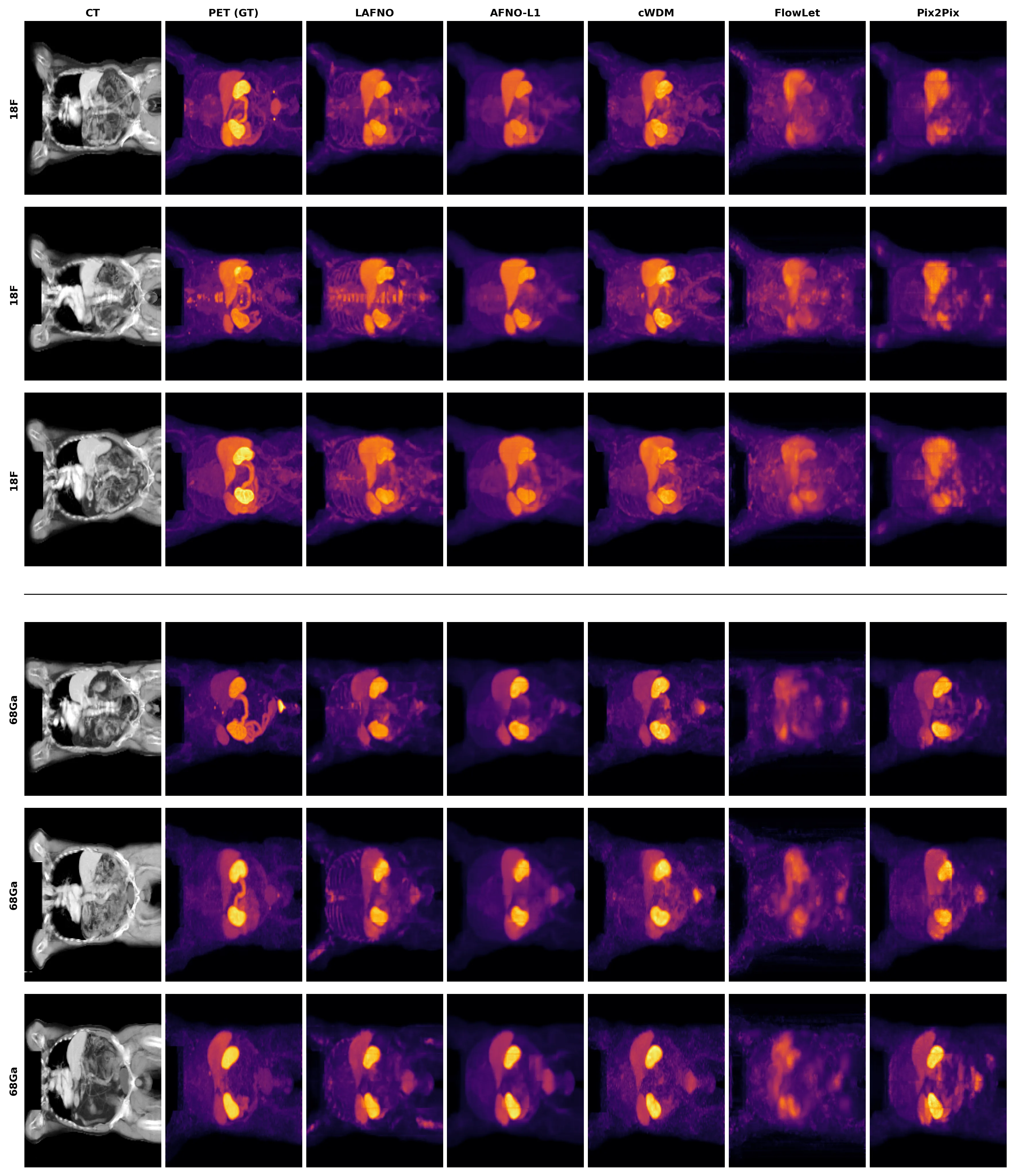
A tumor in a scan is like a tiny bright dot on a vast dark canvas. That dot is what the doctor needs, not a perfectly smooth background. But when a neural network learns to turn a regular CT scan into something resembling a PET scan, it often tries to make everything look nice, and ends up smearing that dot.
The trouble is that tumor cells make up only a minuscule fraction of the body's total volume. If you judge quality by the whole image, the model can score high while still missing the faint glow of cancer. The authors of this new study propose adding two simple signals to the network: one shows how heterogeneous the tissue texture is, the other how strong the density contrasts are. These signals are cheap to compute from CT and help the model focus on suspicious areas.
The model is called LAFNO. It was trained on real patient data from prostate cancer cases, where tumors light up thanks to the radioactive tracer PSMA. In tests, LAFNO cut the error in estimating overall tumor activity by nearly half compared to previous methods. The accuracy of reproducing tumor texture also improved — important for downstream automated analysis.
For now, reproducing the tissue around the tumor remains unstable and depends on the tracer type. But the approach itself, hinting to the network where to look using simple physical features, could be useful in other medical imaging tasks.
Original abstract
as written by the authors arXiv:2608.10429v1
Deep learning models that synthesize PET from CT or MRI can reduce patient dose and scanner demand, but are typically optimized with global losses such as L1 or mean squared error (MSE) that treat all voxels similarly. In whole-body PSMA-PET, tumor voxels occupy only a small fraction of the volume, yet carry the clinically relevant activity signal; as a result, models can achieve high structural similarity index measure (SSIM) and peak signal-to-noise ratio (PSNR) while still underestimating lesion activity or failing to preserve tumor-specific structure. Radiomics provides biologically meaningful descriptors of tumor intensity and texture, but direct radiomics conditioning is time-consuming because it requires feature extraction from delineated lesion regions. We propose LAFNO, a Lesion-Aware Adaptive Fourier Neural Operator for CT-to-PSMA-PET synthesis that replaces high-dimensional radiomics conditioning with two efficient CT-derived proxy channels. Motivated by radiomics analysis of PSMA-avid tumor core and peritumoral regions, LAFNO uses a contrast proxy for local density variation and a disorder proxy for local texture heterogeneity, both injected into the model bottleneck. LAFNO combines whole-volume reconstruction with lesion-level total lesion activity (TLA), tumor-core contrast, and peritumoral supervision. We evaluated LAFNO against four baseline architectures on the TCIA PSMA-PET-CT-Lesions dataset. LAFNO remained competitive on whole-volume image quality, achieving SSIM of 0.960 and 0.938 for 18F- and 68Ga-PSMA, respectively, while reducing per-patient TLA error to 48.3% and 64.0% for 18F- and 68Ga-PSMA, respectively, and achieving the highest tumor-core radiomics reproducibility across all feature classes for both tracers. Peritumoral reproducibility remained tracer-dependent, indicating that biological fidelity in synthetic PSMA-PET remains challenging.
The radioactive tracer PSMA used in PET specifically latches onto a protein abundant on the surface of prostate cancer cells, making the tumor 'glow' in the scan.