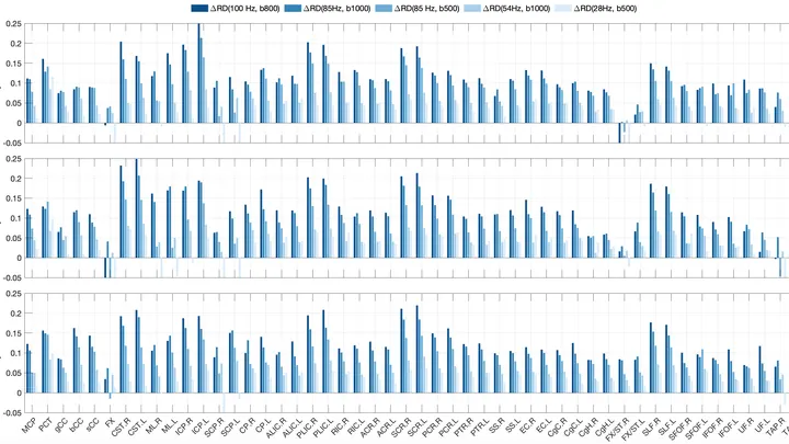
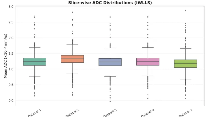
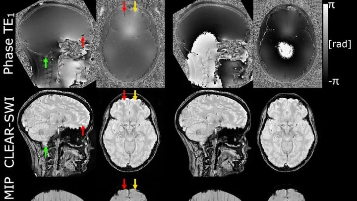
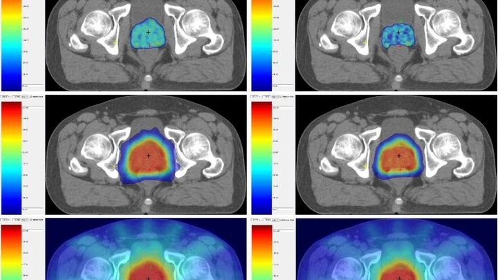
Multiparametric MRI (mpMRI) of the prostate combines several sequences: T2-weighted images show anatomy, diffusion-weighted images reveal cell density based on water molecule mobility, and dynamic contrast enhancement shows blood supply. Combined analysis allows distinguishing malignant tumors from benign changes with high probability.
History
The roots of MRI go back to 1938, when Isidor Rabi discovered nuclear magnetic resonance. In 1973, Paul Lauterbur obtained the first MR image, and in 1977, Peter Mansfield developed echo-planar imaging, speeding up scanning. For the prostate, the first studies appeared in the 1980s, but it was not until the mid-2000s that the multiparametric approach took shape. In 2012, the European Society of Urogenital Radiology (ESUR) introduced the standardized PI-RADS reporting system, revised in 2015 and 2019. Today, mpMRI is a key step in prostate cancerdiagnosis, reducing the number of unnecessary biopsies and improving the detection of aggressive forms.
How it works
A high-strength magnetic field (1.5 or 3 T) aligns the nuclei of hydrogen atoms. A radiofrequency pulse excites them, after which they relax, emitting signals that are encoded by gradient fields and converted into an image. The T2-weighted sequence reflects differences in transverse relaxation time, showing anatomy: the normal peripheral zone is bright, cancer is dark. Diffusion-weighted imaging at different b-values is sensitive to Brownian motion of water; by calculating ADC, a map is obtained where areas of restricted diffusion (high cell density, typical of cancer) appear dark. Dynamic contrast enhancement tracks contrast uptake and washout, revealing zones of neoangiogenesis — tumors 'light up' in the early phases. Combined assessment using the PI-RADS scale minimizes subjectivity.
In practice
It is used for early detection of prostate cancer, biopsy planning, and monitoring disease progression, replacing random tissue sampling with a targeted approach.
Fun fact
Prostate MRI can detect cancer the size of a pea, which is impossible to feel with a finger, and helps avoid unnecessary biopsies in 30% of patients.
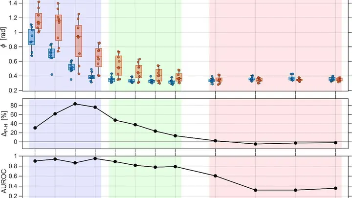
A bowl of jelly trembles but holds its shape when given a gentle shake. Thick syrup, in response to the same motion, sloshes and spreads. A healthy liver is like jelly, but an inflamed one at low frequencies becomes more like syrup.
Researchers built a device that shakes the liver at twelve different frequencies — from five to eighty oscillations per second.
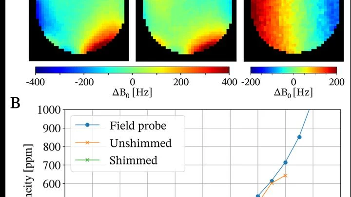
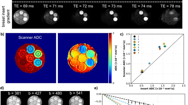
An MRI scanner is a big magnet that aligns hydrogen atoms in the body and then picks up their response signal. Such machines usually take up an entire room and cost as much as a nice house. Engineers from several labs decided to build a scanner that fits in a suitcase and released all the plans as open source.
An MRI scanner is a big magnet that aligns hydrogen atoms in the body and then picks up their response signal. Such machines usually take up an entire room and cost as much as a nice house. Engineers from several labs decided to build a scanner that fits in a suitcase and released all the plans as open source.
Inside an MRI magnet, current flows through a closed superconducting loop, like water circling in a bucket after you give it a spin, it just keeps going on its own. To change that current, you heat the loop with a small heater: it stops being superconducting, becomes an ordinary resistor, and then an external source can 'top up' or 'draw off' current.
A tumor in a scan is like a tiny bright dot on a vast dark canvas. That dot is what the doctor needs, not a perfectly smooth background. But when a neural network learns to turn a regular CT scan into something resembling a PET scan, it often tries to make everything look nice, and ends up smearing that dot.
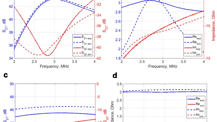
The coil in an MRI picks up radio signals from hydrogen atoms, like an antenna catching a distant radio station. But the signal is weak, and if the amplifier itself is noisy, the image comes out grainy.
The coil in an MRI picks up radio signals from hydrogen atoms, like an antenna catching a distant radio station. But the signal is weak, and if the amplifier itself is noisy, the image comes out grainy.
Water inside nerve fibers doesn't move like it does in a glass: it constantly bumps into the walls of axons (the long projections of nerve cells). If you make the pause between MRI radio pulses very short, water doesn't have time to travel far, and its movement reflects the tightness of the surrounding space.
Prostate MRI scans often look like photos taken through a dirty window: noise, blurred edges, curvature from field imperfections. Scientists checked what happens if you 'wash' that window: remove the excess, correct the distortions, and accurately align different types of scans.
A proton bunch flies into a plasma cloud, and leaves behind an invisible trail: an electric wave. This wave can pick up other particles and accelerate them to enormous energies. But controlling it is tricky: there are too many details in plasma behavior.
Dozens of copper coils in an MRI scanner pick up the faint response from tissues, like microphones trying to catch a whisper in a noisy hall. Each microphone drags a thick bundle of wires behind it. The wires press on each other, create electrical interference, heat up, and add risk for the patient.
The powerful magnet of an MRI scanner induces weak electric currents in tissues. Their phase behaves like a compass needle trembling with every gust of wind — the slightest disturbance turns the map into a ripple, where instead of small vessels or iron deposits, all you see is noise.
In a small Petri dish sits a droplet of liquid the size of a pinhead. An ordinary scanner wouldn't even notice it: the signal is too weak. But bring a tiny magnetic field sensor close to the droplet, and the picture changes.
As a person breathes, the liver glides smoothly up and down. When treating a tumor with radiation, this motion must be accounted for: the beam must hit the target, not healthy tissue. Usually, breathing is "captured" using special computed tomography (CT), taking a series of frames over one breath cycle.
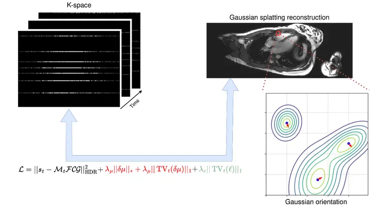
A photographer shoots a runner with a long exposure and gets a blurry silhouette. That's roughly how a standard MRI scanner "sees" moving organs: the heart contracts, the lungs breathe, and the image smears. To avoid this, patients are asked to freeze and hold their breath. But for a child or someone short of breath, that's nearly impossible.
The method called GRASP tackles the problem differently.
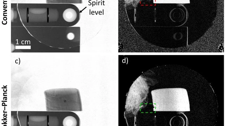
Ultrasound isn't just for baby pictures on a screen. It can scan muscles and tendons, but there's a catch: bones. To sound waves, they act like mirrors to light. The reflection is so strong that nothing behind them is visible.
The scene is lit by spotlights, but the most interesting action happens in the shadows — where molecules invisible to the eye are hiding. The new camera picks up the heat of these "invisible" objects and turns it into ordinary light.
A proofreader checks a manuscript before it goes to print. They don't know every possible mistake, but they know exactly what flawless text looks like. Spotting a strange symbol or a blurred word, they immediately circle it in red. That's roughly how the new program for checking medical images works.
Your arm lifts a cup from the shelf — a familiar motion you don't notice until muscles tire. Then the trajectory changes: the elbow shifts slightly, the hand trembles. A doctor usually catches these changes by hand, feeling the patient's muscles.
When you need to see inside the brain, you usually lie in a tight tube of an MRI scanner or get zapped with X-rays. Both methods are costly, bulky, and not always available.
A flashlight spreads light in a broad beam, while a laser cuts a narrow path and picks out the finest details. In magnetic resonance imaging (MRI), the gradient, a rapid change in the magnetic field along the body, plays the role of that laser.
To peek inside a muscle, it is illuminated with radio waves in a strong magnetic field: that's how MRI works. Water in cells and water in blood respond differently, but a standard scanner doesn't tell them apart: it produces an averaged image where the signal from capillaries is superimposed on the signal from muscle fibers.
Imagine a water park with many pools connected by narrow passages. Visitors, water molecules, splash around in each pool, but occasionally move to the next one. In a brain tumor, these "passages" are protein channels in cell membranes. Their abundance determines how quickly water moves between cells.
An X-ray machine with gratings takes not one but three pictures at once: a regular image, a map of beam deflections invisible to the eye, and an image of tiny structures that scatter light. Together, they help spot, for example, emphysema or pulmonary fibrosis. But there's a catch: to separate these three layers, you need a clever mathematical trick.
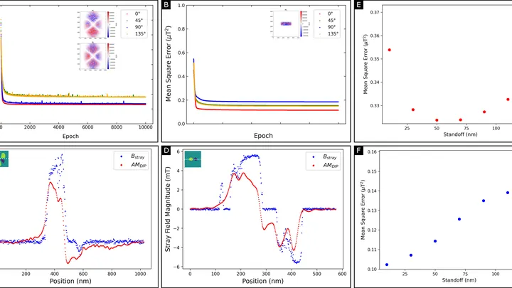
A magnetic film just tens of atoms thick stores information in tiny magnetic vortices. But you can't look inside and see these vortices directly: the instrument only senses the weak magnetic field around the film. Reconstructing the magnetization pattern from such a field is like guessing the shape of an invisible relief from its shadow on a wall.
Two specialists look at the same breast image. One is an experienced doctor, the other is an artificial intelligence program. Sometimes their opinions match, sometimes they diverge. How often they agree determines how accurately they can together distinguish healthy tissue from a tumor.
A surgeon removes a breast tumor, but how do you know no cancer cells are left at the edge? Right now, the tissue goes to a lab, and the answer comes back days later. If the margin is not clean, a second surgery is needed. The new method promises to give the answer right in the operating room, while the patient is still on the table.
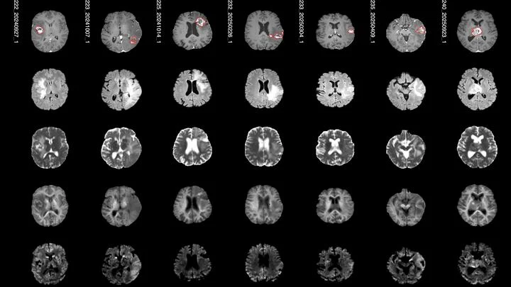
When a person's brain tumor is irradiated, some of the energy also hits healthy cells. A few months later, MRI scans of some patients show bright spots — places where tissue has changed due to radiation. Doctors call them "radiation-induced contrast enhancements." These spots aren't always dangerous, but they show where the brain took the hardest hit.
When a doctor irradiates a tumor, they try to spare nearby healthy organs. In the past, the standard approach was to give many small doses of radiation, like watering a lawn with a fine-spray watering can. Now, hypofractionation is increasingly used: fewer sessions, but larger doses, like a high-pressure hose. The question is how to tell whether the neighboring garden beds got overwatered.
A weak magnet in an MRI machine is like a dim bulb in a dark room: you get a picture, but details drown in grain. These scanners operate at fields below 0.1 tesla (a typical hospital machine runs at 3 tesla, 30 times stronger), yet they're lightweight, inexpensive, and don't require a special room. The catch: noise eats away at quality.
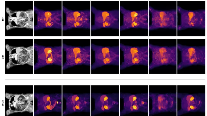
The radioactive isotope terbium-149 acts like a tiny homing bomb: it latches onto cancer cells and detonates with alpha particles that destroy the tumor from within, barely touching healthy tissue. But this bomb has a quirk: it also glows with positrons, and that glow can be captured by a special camera.
Fireflies flash in a dark forest: you see each flash, but not the flight. A quantum gas microscope can show individual cold atoms, like those fireflies. But until now, it couldn't tell you how fast they're moving.
Now scientists have found a way to fix that.
The camera that hunts for tumors works like a highly sensitive camera: it catches the faint glow from a substance that accumulates in diseased cells. In conventional machines, the light is born in a crystal — a solid piece that resembles glass. But a crystal doesn't always convey exactly where the light flashed, so the image comes out slightly blurry.
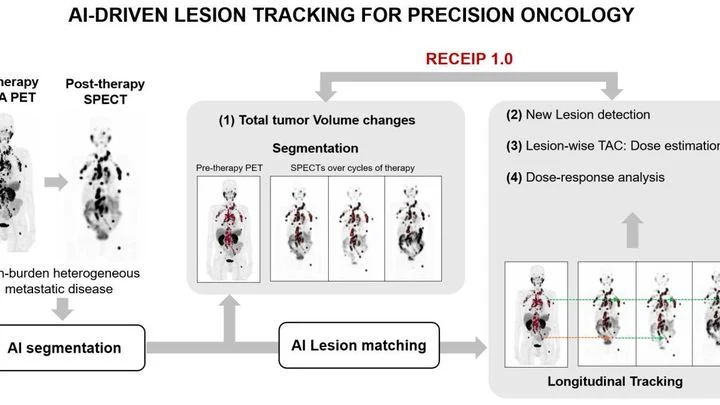
When treating prostate cancer, doctors often see not one tumor but many lesions in bones and lymph nodes. Each lesion can respond to therapy differently: one shrinks, another stays the same, a third grows. Previously, the disease was assessed as a whole, like judging the weather by the average temperature over a year.
In a hospital, blood from a finger prick usually goes to the lab, and the result comes back the next day. The new method promises to cut that wait to a few minutes. Instead of catching light reflected off a sample, it reads the electrical voltage that arises at the boundary between two media, like the faint tingle you feel when you touch a battery with wet fingers.
The placenta is like a bustling market where oxygen passes from mother to child. If the stalls are neatly arranged and goods are delivered quickly, everyone gets enough. But when the aisles get clogged, traffic jams start: some places are crowded, others empty.
An MRI scanner is like a camera that photographs your insides, but very slowly. The patient has to lie still for several minutes while the heart keeps beating. If the person moves or the rhythm goes off, the image blurs. Doctors then have to repeat the scan, wasting time and nerves.
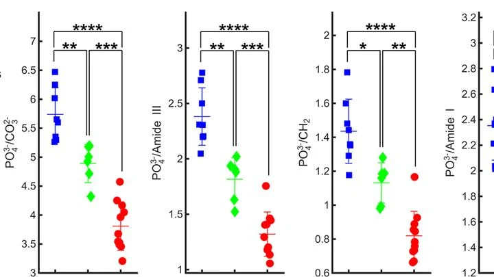
A finger is illuminated with a laser beam, and some of the light returns after traveling inside. That's how Raman spectroscopy works: molecules in bone and skin alter the color of the reflected beam differently, leaving their chemical fingerprints. But skin, muscle, and fat are noisy: their signal drowns out the bone's, like a crowd's chatter overpowering a single singer.
PET/CT is like a map where cancer cells light up as bright spots. But what if the map is blank and the disease is still hiding somewhere? In 132 men with recurrent prostate cancer, doctors found no glowing lesions, but they decided to study the texture of the scan itself, like an experienced tracker noticing bent grass where an animal has passed.
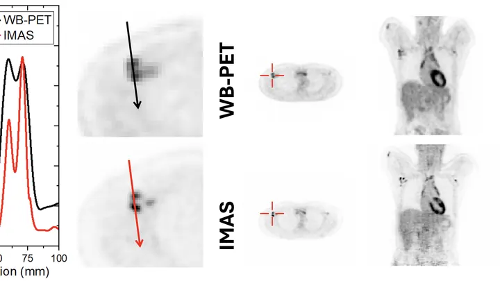
A standard PET scanner is like a camera with a narrow lens: to capture the whole body, you have to move it back and forth and take many shots. The new IMAS tomograph is a wide-angle camera that covers 71 centimeters of the body at once. The patient lies still, and the machine collects an image from head to thighs in a single pass.