Seeing the Heart's Flicker in Two Mirrors at Once express
The heart doesn't beat like a metronome but like a jazz drummer: sometimes it stumbles, skips beats, or races. Atrial fibrillation is one of the most insidious glitches: the atria quiver instead of contracting, and blood can pool, forming clots. Doctors catch this glitch on an ECG — a recording of the heart's electrical signals. But the signal is finicky: it looks different in different people, on different machines, in different hospitals.
Typical algorithms look at an ECG either as a line over time or as a set of frequencies — a spectrum. The first view catches pauses and spikes; the second, rhythmic patterns. But on their own, they're like two witnesses who saw an incident from different angles. The authors combined these views into one algorithm. Two "translators"—time and frequency — work as a pair: each refines what the other noticed, and together they give a more reliable picture.
The second key is contrastive learning. The algorithm is shown pairs of ECGs: "these two are from the same condition, and these are from different ones." So it learns not just to memorize examples but to understand what distinguishes a healthy rhythm from a sick one. It's like teaching a child to tell a cat from a dog by showing not just pictures but also explaining: "see, this one's ears are perky, and that one's tail is up."
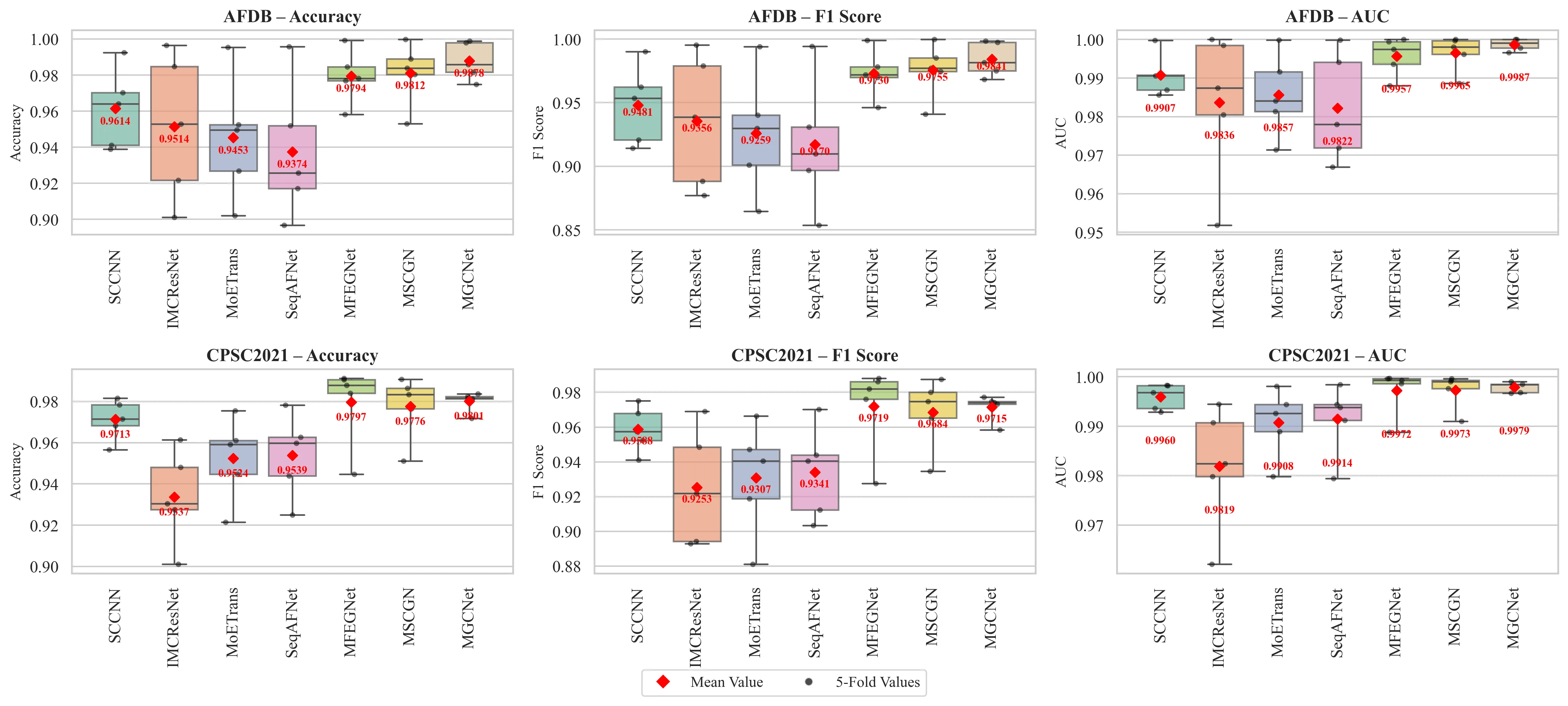
Testing showed the algorithm works reliably even on data from other clinics, recorded with different devices. That's important because in real life, an ECG from a neighborhood clinic and one from a cardiology center look different. The method is also fast: it can run right on a small device at the patient's bedside, without waiting for a powerful computer.
So the two witnesses who used to argue now cross-check their accounts and together see what each missed alone. The heart stops being a puzzle to the algorithm, and the doctor gains a reliable assistant that never tires and never gets distracted.
A human heart beats about three billion times over a lifetime, like a metronome ticking nonstop for 80 years.