A CT (computed tomography) scan is a medical imaging technique that combines multiple X-ray images taken from different angles to create detailed cross-sectional images of the body. Unlike a single X-ray, which compresses three-dimensional structures into a flat image, a CT scan reconstructs the internal anatomy slice by slice, allowing doctors to see organs, bones, and tissues in high resolution. It is essential for diagnosingtumors, internal injuries, and vascular diseases.
History
The first CT scanner was developed by Godfrey Hounsfield and Allan Cormack in the early 1970s, who shared the 1979 Nobel Prize in Physiology or Medicine for their work. The first clinical scan was performed in 1971.
How it works
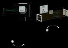
The CT scanner rotates an X-ray tube and detectors around the patient, capturing hundreds of projection images. A computer algorithm, typically using filtered back projection or iterative reconstruction, solves the inverse problem to compute the X-ray attenuation coefficient for each voxel (3D pixel). The result is a stack of cross-sectional images that can be rendered into 3D models.
In practice
CT scans are used in emergency medicine for rapid trauma assessment, in oncology for tumor detection and staging, and in cardiology for coronary artery imaging.
Fun fact
A modern CT scan can image the entire body in less than a minute, but it delivers a radiation dose equivalent to several hundred chest X-rays.
In a dark room, a sunbeam passes through a pinprick in cardboard, and an upside-down image of the street appears on the wall. A pinhole camera needs no lenses: the tiny hole itself selects the rays coming from each point of the scene. Scientists at the ESRF applied this principle not to light but to X-rays, and made the camera see not shape but chemical composition.
A doctor looks at a screen and sees two images of the same part of the body. One shows where blood is rich in oxygen, the other shows how fast it rushes through the finest vessels. Previously, these images were made by different devices, and combining them was difficult. Now engineers have built a device that takes both images simultaneously.
A low-dose X-ray image looks like a view through a fogged-up window: outlines are there, but details drown in a hazy veil. Doctors need such images during radiation therapy — to aim the beam precisely at the tumor and spare healthy tissue. But saving on radiation comes at the cost of noise, streaks, and artifacts that obscure what matters.
Ordinary AI looks at medical images like puzzles made of unfamiliar pieces: it considers any two unmatched pictures completely different. It's like trying to tell twins apart without knowing they're related. In medicine, however, patients with the same condition look nearly identical on MRI, and the algorithm mistakes 'relatives'.
The data from a single CT scan is comparable to several feature-length films. TCAM-Diff bypasses this bottleneck: the program compresses the 3D scene into three shadow projections — like a sculptor reconstructing a solid statue from silhouettes on three walls.
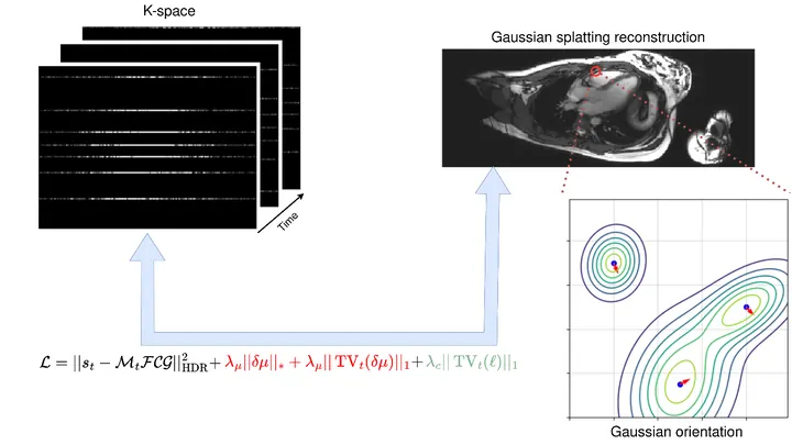
A photographer shoots a runner with a long exposure and gets a blurry silhouette. That's roughly how a standard MRI scanner "sees" moving organs: the heart contracts, the lungs breathe, and the image smears. To avoid this, patients are asked to freeze and hold their breath. But for a child or someone short of breath, that's nearly impossible.
The method called GRASP tackles the problem differently.
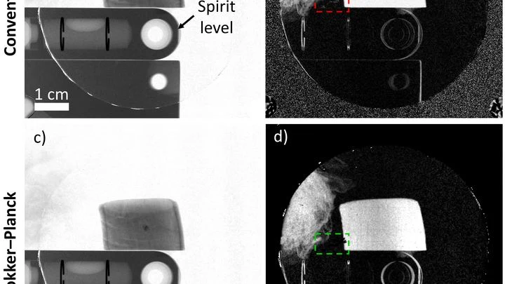
An X-ray machine with gratings takes not one but three pictures at once: a regular image, a map of beam deflections invisible to the eye, and an image of tiny structures that scatter light. Together, they help spot, for example, emphysema or pulmonary fibrosis. But there's a catch: to separate these three layers, you need a clever mathematical trick.
A tomograph is like a photographer who walks around an object and takes pictures from different sides. Usually, it snaps at regular intervals and uses the same amount of light for each shot. But if you need to shoot quickly and not waste extra film, you have to choose: which angles matter more and where to direct more light.
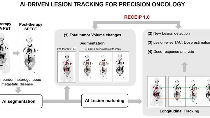
When treating prostate cancer, doctors often see not one tumor but many lesions in bones and lymph nodes. Each lesion can respond to therapy differently: one shrinks, another stays the same, a third grows. Previously, the disease was assessed as a whole, like judging the weather by the average temperature over a year.
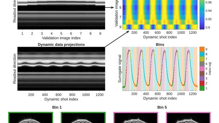
An MRI scanner is like a camera that photographs your insides, but very slowly. The patient has to lie still for several minutes while the heart keeps beating. If the person moves or the rhythm goes off, the image blurs. Doctors then have to repeat the scan, wasting time and nerves.
A beam of carbon ions strikes the tumor precisely, like a surgeon who can only see the skin. Inside the body, the beam stops, and exactly where used to be a mystery. Now, helium is mixed into the same beam: it passes through the body like an X-ray, and its trail shows where the carbon went.
A standard MRI machine is a huge magnet that you can't move, plus a powerful computer that turns signals from the body into a picture. In recent years, portable scanners with weaker fields have appeared: they are light, cheap, and can work in a field hospital or an ambulance.
A photographer snaps a runner: to avoid a blurry shot, a short shutter speed is needed. An MRI scanner is like that photographer, but its 'shutter speed' is long — by the time it captures, the muscle has already changed. That's why dynamic MRI (a series of volumetric frames in motion) is a tough challenge: data are scarce, and motion doesn't wait.
Radioactive tracers are like a courier with a beacon: you inject it into the body, and it highlights the target organ on the scan. But this courier comes with strict rules: you can't send it often, it quickly 'gets tired,' and it costs a lot. Doctors have long dreamed of a way to see an organ without radiation.
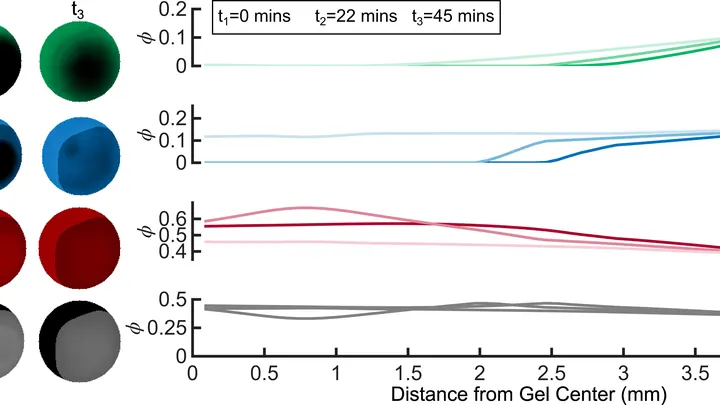
In winter, puddles freeze, and ice cracks the asphalt. Inside a living cell, freezing does something similar: ice crystals tear its membrane. To prevent this, the cell is soaked in a special solution, like antifreeze for a car. But precision is key: too little solution and ice still forms; too much and the solution itself poisons the cell.
Cells breathe. They need oxygen, and they take it from the blood flowing through the tiniest vessels. If blood flow is weak or oxygen is scarce, cells start to suffocate. Doctors need to see this as early as possible. But how do you look inside living tissue without cutting it open?
The new device does it in two ways at once.
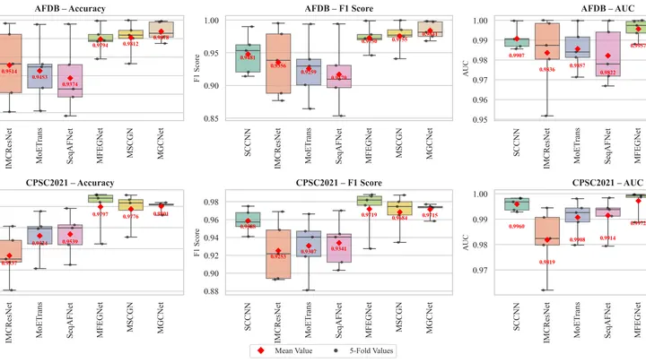
The heart doesn't beat like a metronome but like a jazz drummer: sometimes it stumbles, skips beats, or races. Atrial fibrillation is one of the most insidious glitches: the atria quiver instead of contracting, and blood can pool, forming clots. Doctors catch this glitch on an ECG — a recording of the heart's electrical signals.