The Radiation Scalpel Gets Sharper express
Radiation therapy is like shooting at a small target with a very large-caliber rifle: even a precise shot hits a large area around it. The thickness of the 'trace'—called the penumbra — is 2–3 millimeters in standard machines. For a tumor in the lung, that's not a big deal, but next to a nerve or a blood vessel, those millimeters decide whether a person stays healthy.
Physicists from the clinic and the university tried an unconventional approach: they lowered the beam energy from 6 to 2.5 megavolts and moved the patient four meters away from the source instead of the usual one. Individually, these steps worsen the beam, but together they produce an unexpected effect — the penumbra shrinks to one millimeter. At the same time, the beam still reaches tumors at a depth of ten centimeters.
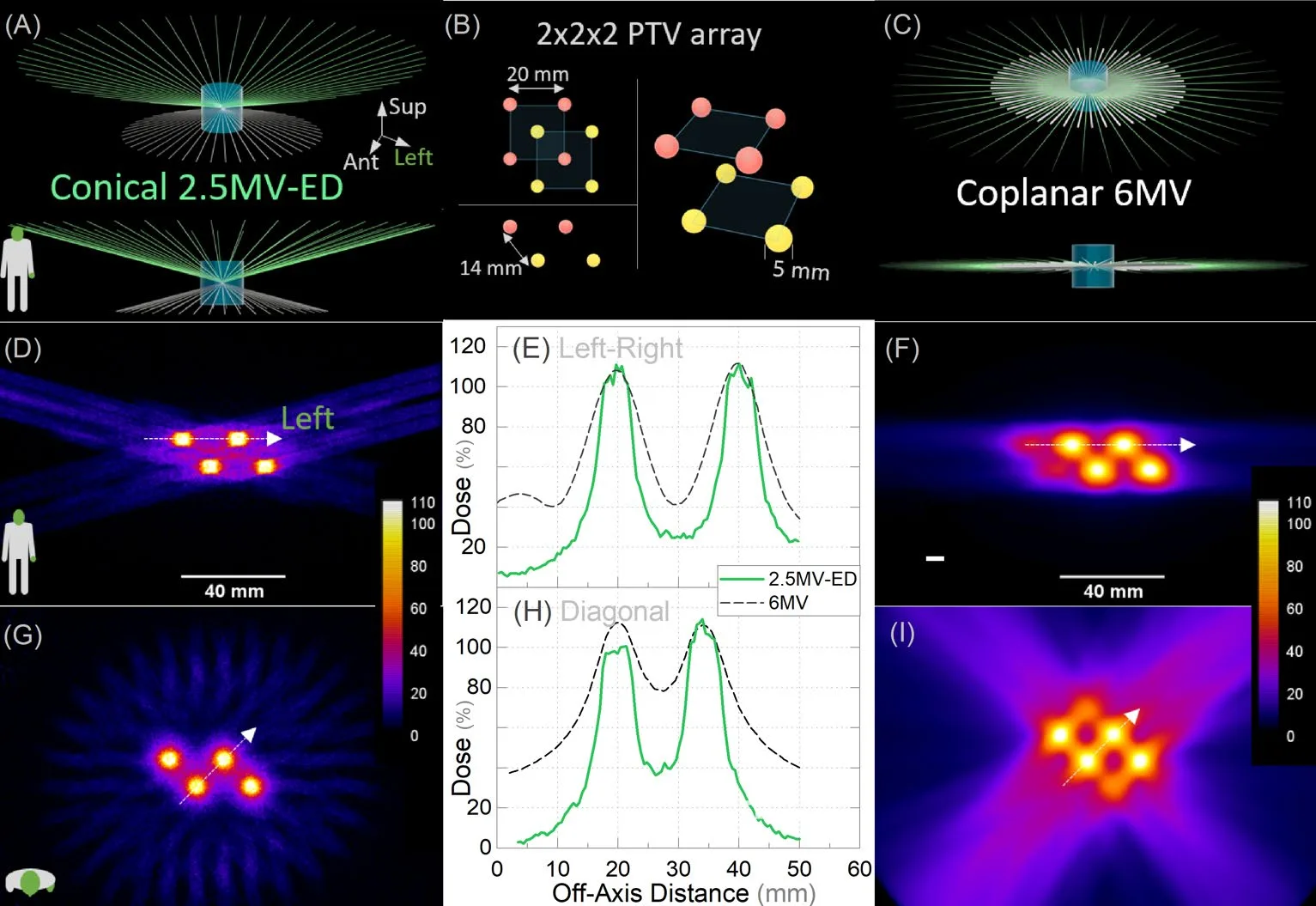
To test the idea, the researchers built a setup where the patient sits upright and the radiation sources are arranged around them in a cone. In a computer model, this scheme gave a 1.7 times more contrast dose distribution than the standard one. In one example, irradiating spheres with a diameter of 5 millimeters inside a tumor, the contrast between 'hot' and 'cold' spots increased from 2.6 to 4.5.
This means a doctor could burn out tiny nodules inside a tumor without touching the healthy tissue between them. For the patient, there is less risk of skin burns and damage to neighboring organs, and for cancer treatment, it's a step toward making the radiation knife a truly surgical instrument.
The penumbra is the distance over which the dose drops from 80% to 20% of the maximum. For the new beam, it's just one millimeter — thinner than two human hairs stacked together.
In science fiction, ray weapons are often depicted as thin light sabers. The new technology makes that image almost real for medicine: the beam can be sharpened to a millimeter edge.