Invisible Proteins Start to Talk express
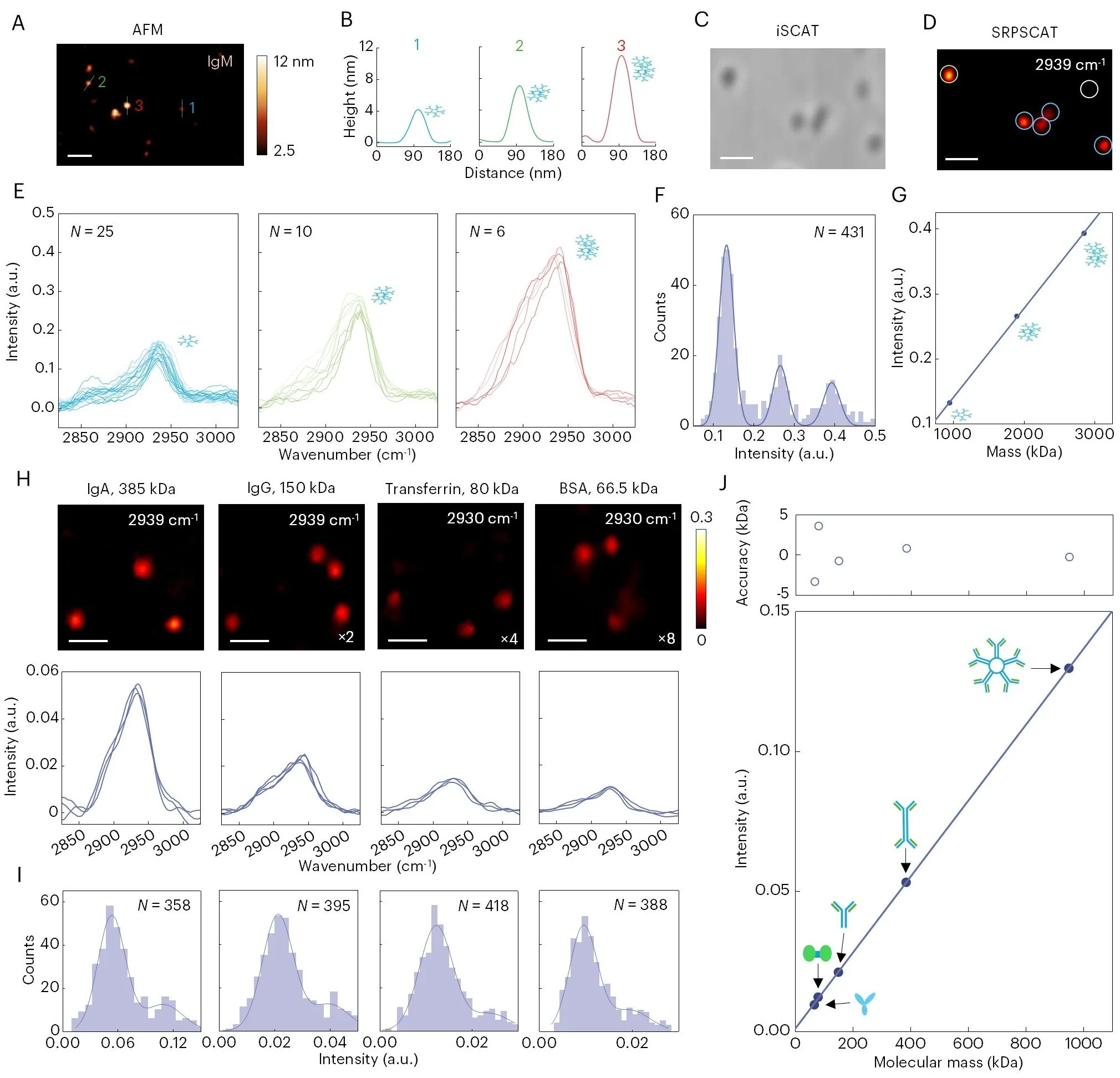
A protein in a cell is like a tightly coiled spring. Its shape determines what it does, but seeing that shape in a single molecule without disturbing it was nearly impossible. The new microscope works differently: it sends a short laser pulse that slightly heats the molecule. The heat makes the molecule vibrate, and these vibrations are like a unique voice that reveals both the protein itself and its state.
The method is called photothermal coded scattering spectroscopy. The laser 'listens' to the vibrations of bonds inside the protein — the very bonds that link atoms in a chain. Each bond vibrates at its own frequency, and the set of frequencies forms a fingerprint, like a chord made of several notes. By comparing 'chords,' scientists can distinguish proteins, determine their mass, and even see how a protein changes shape while working.
Previously, proteins were labeled with glowing dyes, but the label could alter the molecule's behavior, like a heavy ornament on a spring. The new method requires no labels, so proteins remain in their natural state. Scientists have already measured how quickly one protein binds to another and watched an engineered protein switch between two forms, like a key turning in a lock.
Now it will be possible to study proteins in their native environment — inside the cell, not in a test tube. This is a step toward understanding how the molecular machines of life work and toward creating drugs that hit their targets more precisely.
Original abstract
as written by the authors arXiv:2603.25534
Current single molecule methods either rely on fluorescence or lack chemical information. Here we report stimulated Raman photothermal encoded scattering (SRPSCAT) microscopy for quantitative bond-selective imaging of single-biomolecule structures and interactions in native environments. In this approach, scattering of the target molecule is modulated by the deposited energy from stimulated Raman gain and loss processes, thereby encoding vibrational spectroscopic information. Leveraging single-molecule sensitivity of interferometric scattering, SRPSCAT can map single proteins with chemical specificity, determine their mass, and distinguish protein secondary structures based on their Raman fingerprints. Furthermore, single protein binding kinetics are quantified and the conformational dynamics of single de novo designed allosteric proteins are observed. Together, these results highlight the potential of SRPSCAT for label-free structural, functional and dynamic analysis at the single-molecule level.
Proteins vibrate at frequencies up to a billion times per second — faster than a mosquito's wingbeats.