Invisible Proteins Start to Talk express
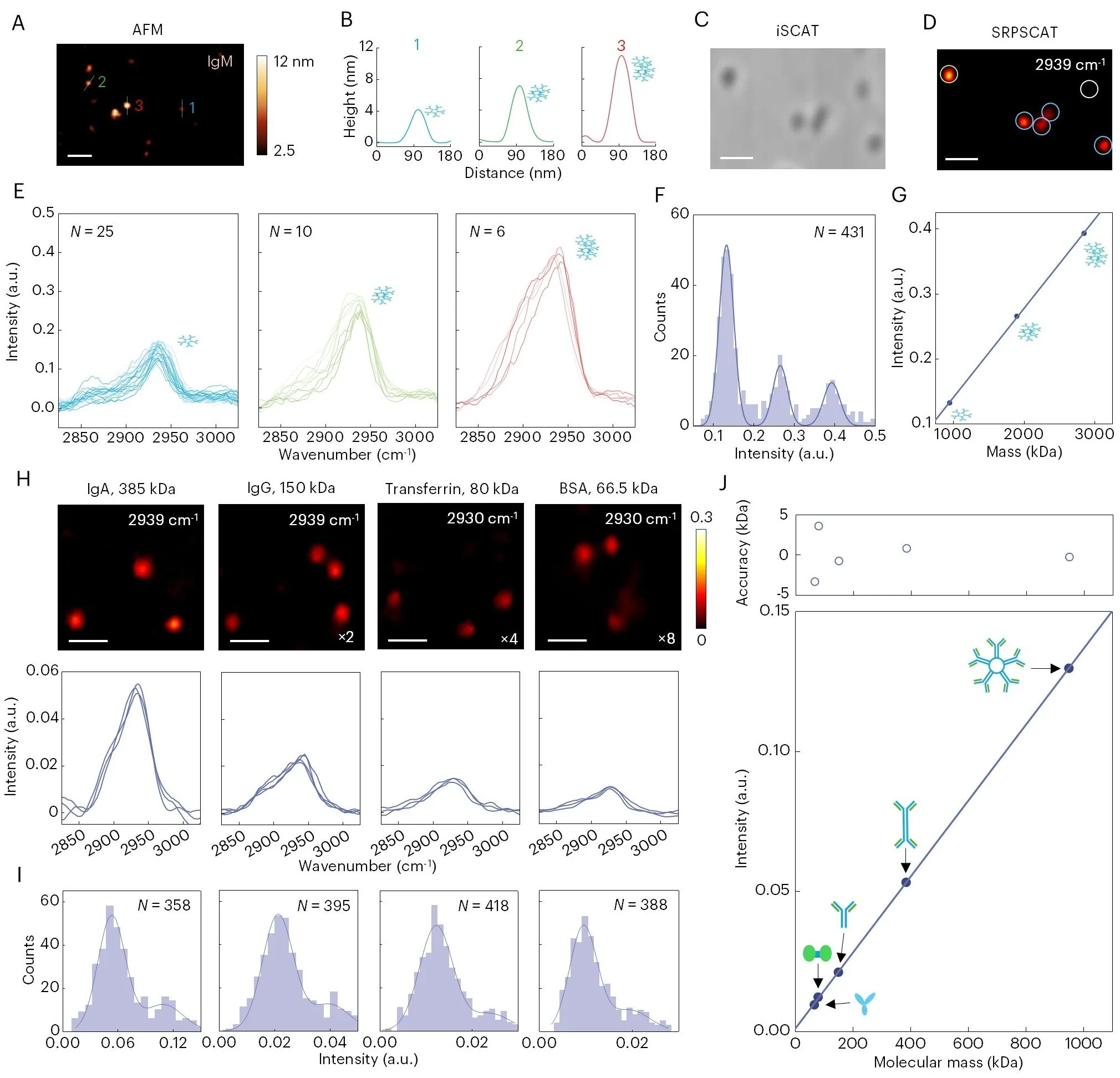
A protein in a cell is like a tightly coiled spring. Its shape determines what it does, but seeing that shape in a single molecule without disturbing it was nearly impossible. The new microscope works differently: it sends a short laser pulse that slightly heats the molecule. The heat makes the molecule vibrate, and these vibrations are like a unique voice that reveals both the protein itself and its state.
The method is called photothermal coded scattering spectroscopy. The laser 'listens' to the vibrations of bonds inside the protein — the very bonds that link atoms in a chain. Each bond vibrates at its own frequency, and the set of frequencies forms a fingerprint, like a chord made of several notes. By comparing 'chords,' scientists can distinguish proteins, determine their mass, and even see how a protein changes shape while working.
Previously, proteins were labeled with glowing dyes, but the label could alter the molecule's behavior, like a heavy ornament on a spring. The new method requires no labels, so proteins remain in their natural state. Scientists have already measured how quickly one protein binds to another and watched an engineered protein switch between two forms, like a key turning in a lock.
Now it will be possible to study proteins in their native environment — inside the cell, not in a test tube. This is a step toward understanding how the molecular machines of life work and toward creating drugs that hit their targets more precisely.
Proteins vibrate at frequencies up to a billion times per second — faster than a mosquito's wingbeats.