How the Robin 'Sees' the Magnetic Field express
Every autumn, a robin weighing as much as a pat of butter lifts off from a branch somewhere near St. Petersburg and flies south toward the Mediterranean, never once checking a map. It is believed that its eye contains a built-in magnetic compass, and the main component of this device is the protein cryptochrome 4a. Blue light triggers an electron transfer within it, giving rise to a radical pair, two molecules with unpaired electrons, whose quantum state depends on the tilt of Earth's magnetic field. The problem is that the quantum lifetime of such a pair lasts only microseconds, while the bird needs a signal that lasts long enough for a nerve cell to notice.
The researchers studied the ErCry4a protein from the European robin using a method that can be compared to flash-freezing a moving mechanism. They irradiated the protein with blue light and then, at various time points, plunged it into heavy water. Where the protein chain was mobile and open, hydrogen atoms were quickly replaced by deuterium; where the structure was rigid, the exchange was slow. This allowed them to capture a 'snapshot' of the protein's shape at each stage: the dark state, the transition to the active state under light, the short-lived semiquinone, and the final, fully reduced state.
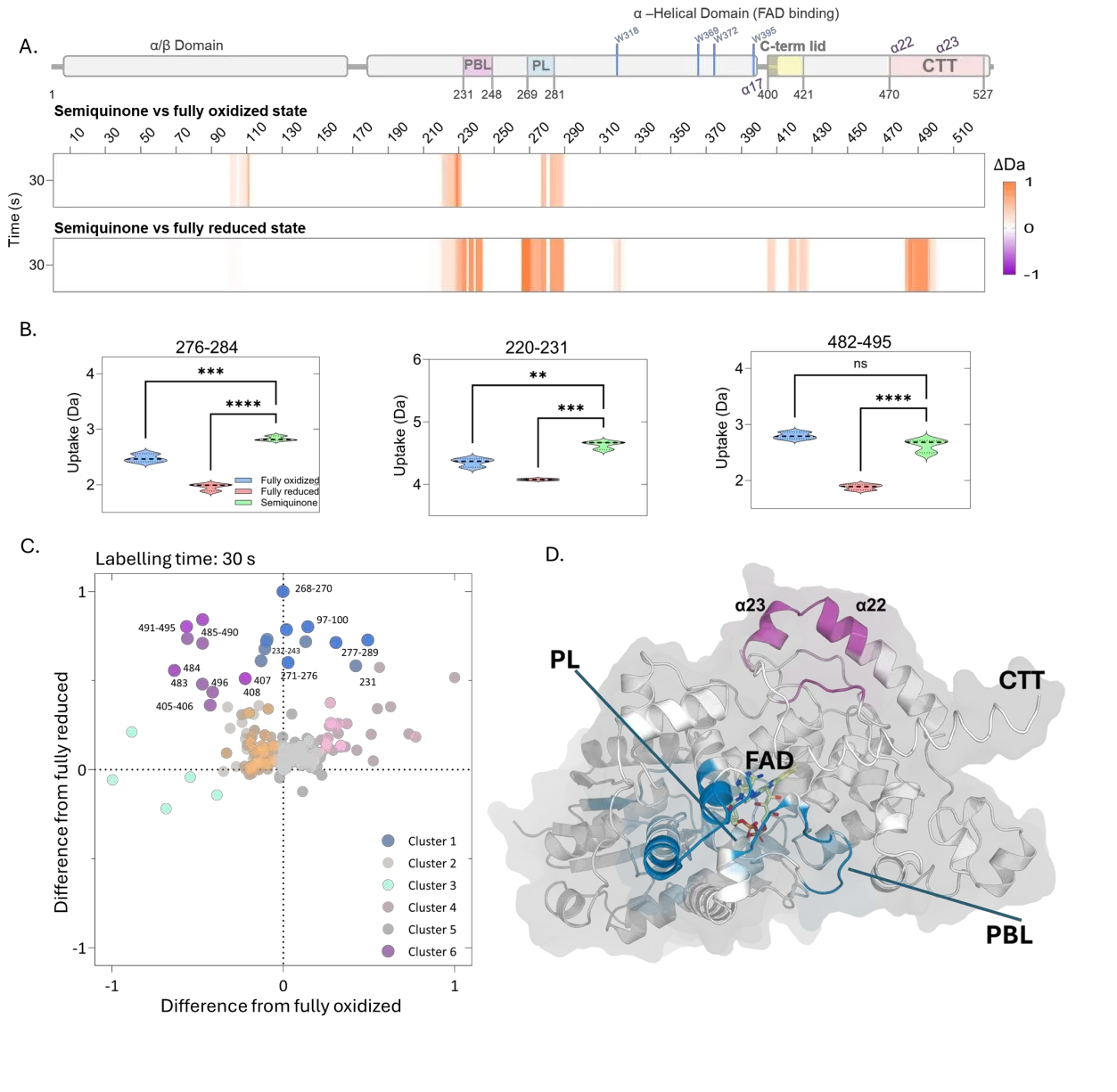
It turned out that the semiquinone behaves not like a bridge but like a distinct entity with its own character. In the fully reduced protein, everything becomes rigid, as if the mechanism were locked in one position. In the semiquinone, by contrast, two important loops, the phosphate-binding loop and the protruding loop, lose stability and become mobile. It is this instability, rather than rigidity, that may serve as a clear signal for the next player in the chain: a mobile loop can touch a partner protein and trigger a cascade that eventually reaches the bird's brain. Thus, a fleeting quantum event gains a long biochemical life.
Original abstract
as written by the authors arXiv:2604.19579
The light-dependent magnetic compass of night-migratory songbirds is widely hypothesized to rely on the radical pair mechanism within retinal cryptochrome. However, bridging the mechanistic gap between microsecond quantum spin dynamics and the long-lived, global protein conformational changes required for cellular signalling remains a formidable challenge. Here, we apply redox state-resolved hydrogen/deuterium-exchange mass spectrometry (HDX-MS) to map the conformational landscape of European robin cryptochrome 4a (ErCry4a) across its photocycle. We reveal that photochemical reduction drives robust, allosteric structural transitions across key functional nodes, including the phosphate-binding loop (PBL), protrusion loop (PL), FAD-proximal helix {\alpha}17, and the C-terminal {\alpha}22/{\alpha}23 network. Crucially, we isolate the structural fingerprint of the transient semiquinone, the presumed signalling species. Rather than acting as a linear structural stepping-stone, the semiquinone exhibits a distinct, non-monotonic conformational signature characterized by a transient destabilization of the PBL and PL, contrasting sharply with the global rigidification observed in the fully reduced state. These findings establish the semiquinone as a structurally unique and functionally competent biological entity. Our results provide direct biophysical evidence for a dedicated, high-fidelity structural signalling cascade, detailing how localized quantum-level photochemistry is translated into the precise conformational dynamics required for animal navigation.
A robin can detect the inclination of the magnetic field with an accuracy better than five degrees — roughly like being able to tell, with your eyes closed, which way a compass needle points by the faint warmth on your skin.