How a Low-Dose X-Ray Becomes a Sharp Image express
A low-dose X-ray image looks like a view through a fogged-up window: outlines are there, but details drown in a hazy veil. Doctors need such images during radiation therapy — to aim the beam precisely at the tumor and spare healthy tissue. But saving on radiation comes at the cost of noise, streaks, and artifacts that obscure what matters.
The authors built a neural network that acts like a patient restorer. It takes a corrupted image and, step by step, removes defects, revealing clear structure. At its core is a diffusion model, an algorithm that learns from pairs of 'blurry image, clean image' and then recovers details even from very noisy data. It's like a program trained to guess the original from a blurred photo.
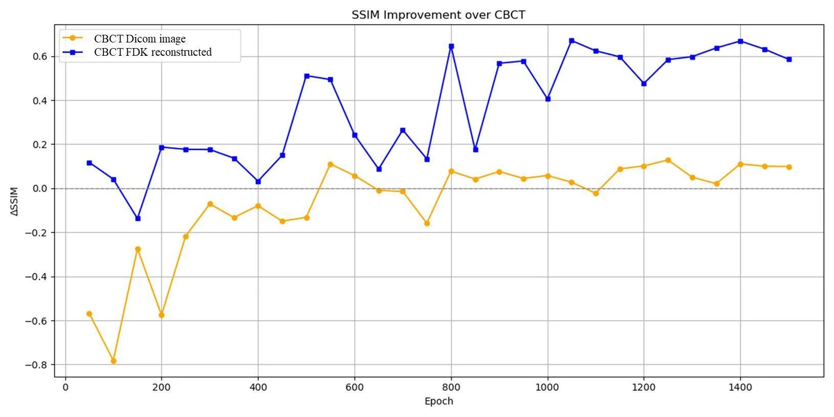
The network was trained on images from patients undergoing radiation therapy, and the synthesized CT was checked against the real thing. It turned out that a physically meaningful data representation is not just a convenient abstraction but a real way to improve accuracy. For the clinic, this means less radiation during the control phase while maintaining high planning quality. Thus, the foggy glass becomes transparent, and the doctor sees every millimeter of tissue.
Original abstract
as written by the authors arXiv:2608.08919v1
During standard radiotherapy planning, repeated CT acquisitions are often required for patient registration, verification, and adaptive planning, resulting in increased cumulative X-ray dose. To mitigate this, low-dose cone-beam CT (CBCT) is routinely acquired during treatment delivery. However, CBCT image quality remains insufficient for accurate dose calculation and adaptive radiotherapy planning due to increased scatter, noise, beam hardening, and reconstruction related artifacts. This study develops a supervised deep learning based CBCT to CT synthesis framework using a conditional denoising diffusion probabilistic model (DDPM), where the generation of a CT-based planning for accurate positioning and dose calculation is obtained using generative models with low dose CBCT imaging. Beyond demonstrating CBCT to CT synthesis, the primary objective is to investigate how the representation of CBCT input data, either standard clinical DICOM CBCT images or filtered back-projection (FDK) reconstructions from raw projection data, affects the performance of diffusion based CT synthesis. The overarching aim is to assess whether physics aware CBCT representations better support CT-equivalent image quality while maintaining reduced imaging dose in radiotherapy workflows.
Diffusion models, like the one used here, were originally invented for generating images from text descriptions: they power popular AI art generators. Now the same idea helps clean noise from medical images.