Image reconstruction is the computational process of converting raw detector signals or measurements into a meaningful visual image. In many imaging systems, the detector does not directly capture a picture; instead, it records projections, interference patterns, or other indirect data. Reconstruction algorithms invert the physics of the measurement to recover the original object. This is fundamental in medical imaging (CT, MRI, PET), astronomy, and microscopy, where direct imaging is impossible or impractical.
History
The mathematical foundations were laid by Johann Radon in 1917, who derived the transform that underlies tomographic reconstruction. The first practical medical CT scanner was developed by Godfrey Hounsfield and Allan Cormack in the early 1970s, for which they shared the Nobel Prize in Physiology or Medicine in 1979.
How it works
The process typically involves solving an inverse problem. For example, in computed tomography (CT), X-ray measurements are taken from many angles, and the reconstruction algorithm (often filtered back-projection or iterative methods) uses these projections to compute the spatial distribution of attenuation coefficients. In MRI, raw data are in the form of spatial frequencies (k-space), and a Fourier transform reconstructs the image. Modern techniques often use regularization and machine learning to improve quality and reduce noise or artifacts.
In practice
Image reconstruction is used in virtually all medical imaging modalities, in radio interferometry for astronomy (e.g., the Event Horizon Telescope), and in electron microscopy for structural biology.
Fun fact
The algorithm used in the first CT scanner took hours to reconstruct a single image on a computer that was less powerful than a modern calculator.
A courtroom interpreter catches every word, every breath, every whisper in the corner. They have no right to miss a detail, because the meaning of the whole conversation depends on it. Quantum state tomography works in much the same way: it reconstructs the full quantum state of a system from a set of measurements, leaving no detail out.
A translator at the negotiating table doesn't listen to every word; they grasp the meaning of whole phrases. Electron microscopy poses a similar challenge: you need to figure out the crystal's thickness and its tilt in the beam, and all you have is a diffraction pattern, a mosaic of thousands of bright and dark spots.
A deck of cards in which each card is slightly rotated cannot lie flat: the backs and faces will never align. But if the cards are flexible, they will start to adjust, shifting by fractions of a millimeter to find the perfect match. Something similar happens inside crystals when two thick layers of MoS2 or MoS2 with WS2 are stacked with a twist of 1–2 degrees.
A cartographer receives a sheet with distances between hundreds of unnamed islands. No island is labeled, but the distances repeat, forming triangles and quadrilaterals. From these repetitions, the cartographer reconstructs the map of the archipelago, not knowing which island is which. This is roughly how the new work in molecular spectroscopy works.
Under an electron microscope, a protein looks like a series of blurry shadows — hundreds of thousands of images of the same molecule, but from different angles and with heavy noise. Previously, from these shadows, one first assembled a volumetric 'haze'—a map of how electric charge is distributed inside the protein.
A stenographer is recording a meeting, but halfway through the conversation she runs out of paper. She misses a few lines, then picks up where she left off. Later, she needs to reconstruct the full transcript without rewriting the entire conversation from scratch. That's exactly how the new method URA-SIM works for structured illumination microscopy (SIM).
A smartphone camera captures light with tiny sensors, but by themselves, they can't distinguish colors. So color filters are placed in front of them: red, green, blue, like stained glass in a window. Each sensor sees only its own color, and then software assembles the full picture from these pieces.
A shipping container is a steel safe the size of a room, packed with goods. X-rays struggle to see through it: thick walls and dense cargo block the rays. Enter muons — particles born high in the sky that constantly rain down on Earth. They can pass through even lead, but they veer slightly off course when they encounter something heavy.
A low-dose X-ray image looks like a view through a fogged-up window: outlines are there, but details drown in a hazy veil. Doctors need such images during radiation therapy — to aim the beam precisely at the tumor and spare healthy tissue. But saving on radiation comes at the cost of noise, streaks, and artifacts that obscure what matters.
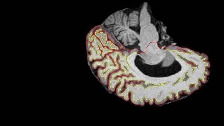
Two scans of the same brain taken at different times are like photographs of the same house before and after renovation. To see what changed, you overlay them and look at the differences. But an MRI scanner is not a perfect camera: it adds its own distortions, as if the photo were taken through slightly warped glass.
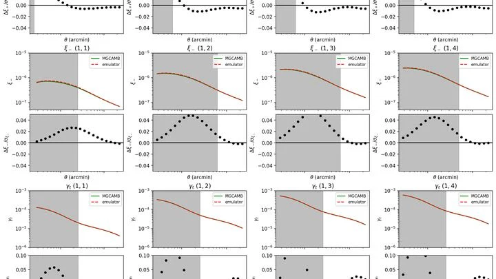
In the vast cosmic ocean, dark matter gathers into clumps and filaments, like plankton in currents. But if gravity doesn't work exactly as Einstein said, these patterns change — barely noticeably, like ripples from an invisible wind. You can spot such ripples by comparing sky maps with computer simulations.
Unlike regular cameras, event-based ones only record moments when light becomes brighter or dimmer. They don't produce snapshots; instead, they send an endless stream of such flashes. Scientists turned this stream into a sharp picture of the Earth while flying over it on a satellite. Each flash is like a piece of a mosaic; together they form a still image.
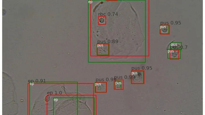
A drop of urine under a microscope isn't a flat puddle but a multi-layered volume. Like a stack of transparent pages: if you focus on one, the neighboring ones blur. The microscope's light only shows one depth layer sharply. To see all the cells, a lab technician had to take a series of snapshots, manually adjusting focus — tedious and slow.
Usually a CT scanner takes many X-ray images from different angles and assembles a 3D picture. But sometimes doctors only scan a small area (say, one lung) and the data cuts off. It's like a chef given a single spoonful of soup and asked to name the exact recipe for the entire pot.
Imagine photographing a statue in dim light. You face a dilemma: take one bright shot or a series of dim frames from different angles. In the first case, quality is limited by photon noise, the graininess that arises from a lack of photons, while in the second, details overlap, making it harder to reconstruct the 3D structure.
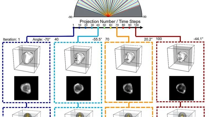
Tomographs don't produce ready-made pictures. They collect numerous 'shadows' — projections of innards from different angles or using various signals — and mathematically reconstruct a 3D picture from them.
Inside a fusion reactor, plasma rages — a gas heated to millions of degrees. There are no direct windows, and cameras and sensors quickly fail. Engineers see only fragments of information: a few sensors at scattered points. It's like trying to grasp a symphony from the sound of three microphones in a vast hall.
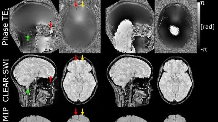
The powerful magnet of an MRI scanner induces weak electric currents in tissues. Their phase behaves like a compass needle trembling with every gust of wind — the slightest disturbance turns the map into a ripple, where instead of small vessels or iron deposits, all you see is noise.
X-ray detectors work like honeycombs: particles flash here and there in hexagonal cells. Conventional algorithms stretch these combs onto a square grid, creating ghostly polarization artifacts. Scientists used a neural network that lives by the rules of the hive — it processes the hexagonal structure directly, preserving the natural pattern.
A single camera shot stores both a normal picture and a hidden map of how light rippled through the scene. Quantum entanglement makes this possible. Pairs of linked photons are generated: one visits the object, its twin takes a detour. Later, measuring the twin one way pulls out the brightness information.
The scene is lit by spotlights, but the most interesting action happens in the shadows — where molecules invisible to the eye are hiding. The new camera picks up the heat of these "invisible" objects and turns it into ordinary light.
A proofreader checks a manuscript before it goes to print. They don't know every possible mistake, but they know exactly what flawless text looks like. Spotting a strange symbol or a blurred word, they immediately circle it in red. That's roughly how the new program for checking medical images works.
Two ultra-dense stars spiral around each other and eventually collide. This dance lasts only moments, but it sends powerful ripples through space — gravitational waves. After the merger, a rapidly spinning blob remains, ringing like a bell, and that ring carries information about what the stars were made of.
A spectrum is like a photograph of a material's inner workings, taken with light. When a beam passes through or reflects off a sample, molecules leave characteristic dark bands, peaks, on that photograph. But if the image is overexposed or veiled in gray, making out details becomes nearly impossible.
An X-ray tomograph is like a carpenter who examines a log from all sides to see where the void is inside. It takes dozens of flat shadow images, rotating the source and detector around the object. Then a 'reconstruction' program glues these shadows into a 3D model. The quickest reconstructor is the FBP algorithm (filtered back projection).
A solar telescope is like an eye trying to spot an ant through a pot of boiling soup. The daytime sky dazzles, the air shimmers, and the image swims. To freeze it, you need adaptive optics — a system that predicts distortions and corrects them hundreds of times per second.
Imagine a river that doesn't flow in an open channel but through the inside of a sponge, with its currents shifting from place to place. That's a good way to think about the brain's glymphatic system: a network of ultra-fine channels around blood vessels, through which cerebrospinal fluid seeps into the tissue and washes away the waste that has piled up during the day.
Cosmic rays are particles from deep space, accelerated to colossal energies. Smashing into the atmosphere, they trigger particle showers with radio waves. These waves are fingerprints, revealing the energy and direction of the intruder.
You taste a fruit smoothie through two different straws. One lets through only the big chunks, the other only the liquid. Individually, the sensations are misleading, but together they give you the key: here, it seems, is banana, and here, strawberry.
Light passing through a turbid medium like milk or living tissue scatters in all directions. Reconstructing where light was absorbed more strongly is like guessing the shape of a fallen object from a puddle on the asphalt. Optical tomography solves this problem, but usually requires large computational resources.
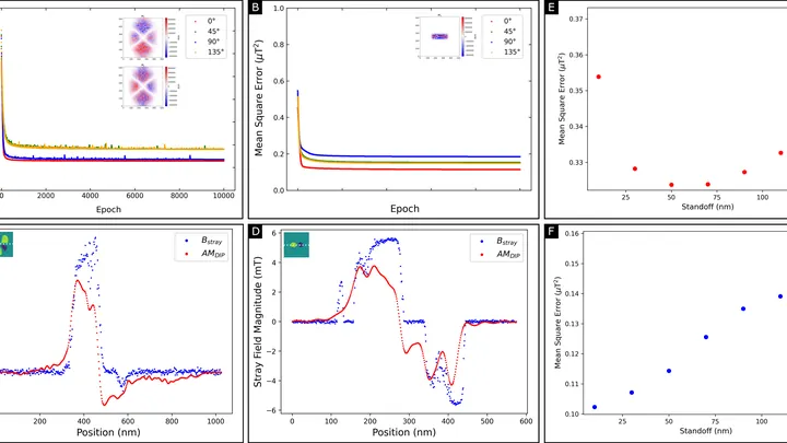
A magnetic film just tens of atoms thick stores information in tiny magnetic vortices. But you can't look inside and see these vortices directly: the instrument only senses the weak magnetic field around the film. Reconstructing the magnetization pattern from such a field is like guessing the shape of an invisible relief from its shadow on a wall.
A tomograph is like a photographer who walks around an object and takes pictures from different sides. Usually, it snaps at regular intervals and uses the same amount of light for each shot. But if you need to shoot quickly and not waste extra film, you have to choose: which angles matter more and where to direct more light.
An ordinary light sensor answers the question "how much light" but not "where did it come from." To do that, you need lenses, mirrors, or entire arrays. In a new study, researchers propose a sensor that determines the direction of a light beam on its own, without any optics.
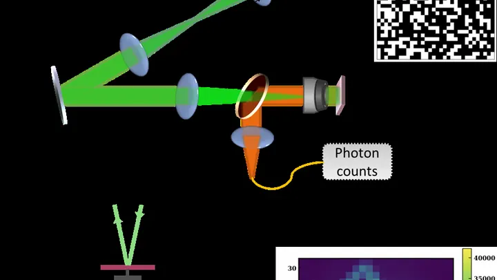
A silicon detector is like photographic film, but instead of light, it senses charged particles. When a particle flies through the silicon, it knocks out electrons, and we see a signal. But how do you look inside the detector itself and see where the electric field is strong and where it's weak? One way is to use a laser.
A giant sphere filled with liquid flashes when an elusive particle, a neutrino, flies through it. Inside the sphere, like fireflies on a Christmas tree, hang thousands of tubes that catch these flashes. To pinpoint exactly where the flash occurred and how much energy arrived, you need to know the position of each tube to within a millimeter.
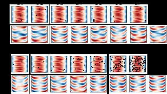
Hunting for a single glowing spot in a dark sample is like trying to find a needle in a haystack: the microscope beam crawls line by line, wasting time and light. Physicists replaced this slow sweep with random light patterns. The sample is illuminated with a broad field, but each time with a new pattern of bright and dark patches, as if random shadows from a grid were projected onto it.
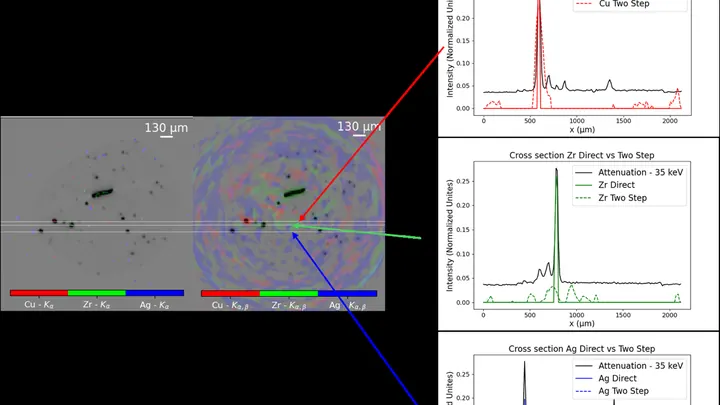
Conventional X-ray fluorescence analysis works like a meticulous scribe: to learn the composition of a picture, it walks over every square centimeter and logs whether lead or copper is present. For a flat sheet, that's tolerable, but for a three-dimensional object, it's torture.
A drummer stays silent, but from the pauses you can tell where he didn’t hit. Similarly, physicists built an image by catching not light, but moments of absolute darkness. The picture emerged from silence.
In the experiment, they shone light from an ordinary lamp onto the object.
An ordinary camera captures light reflected from objects and builds a picture from it. But if the object is hidden behind an obstacle or does not reflect light, radio waves come to the rescue: they pass through walls and fog, and a computer reconstructs the image from scattered signals.
A tiny gold star melts under the beam of an electron microscope, its points slowly rounding off. In the past, such a process could only be seen in snapshots taken with long pauses, and the probing beam itself would alter the sample.