Medical imaging in oncology uses techniques like computed tomography (CT) and positron emission tomography (PET) to detect, characterize, and monitor cancerous tumors and metastases. These methods allow clinicians to visualize the location, size, and metabolic activity of tumors without invasive procedures. The field has evolved from simple anatomical imaging to functional and molecular imaging, enabling earlier detection and more precise treatment planning.
History
The concept of using imaging for cancer detection dates to the early 20th century with X-rays, but modern CT emerged in the 1970s (Hounsfield and Cormack), and PET was developed in the 1950s–1970s. The fusion of PET and CT became standard in the 2000s.
How it works
CT scans use X-rays to create detailed cross-sectional images of the body, revealing tumor size and shape. PET scans inject a radioactive tracer, often fluorodeoxyglucose (FDG), which accumulates in metabolically active cells like cancer cells; the emitted positrons are detected to map metabolic activity. Combining both (PET/CT) overlays functional and anatomical data, helping distinguish benign from malignant lesions and track treatment response.
In practice
Used routinely in oncology for staging, treatment planning, and assessing response to therapy, such as in lung, breast, and prostate cancers.
Fun fact
PET scans can detect tumors as small as a few millimeters, but the resolution is limited by the physics of positron annihilation, not just the scanner.
The electron beam of a microscope usually flies through emptiness, like a courier on an empty highway. Any air molecule is a bump that makes it bounce and lose direction. So before, to examine a sample, you had to freeze it, dry it, or coat it with metal—and still you could only look in a vacuum.
In a dark room, a sunbeam passes through a pinprick in cardboard, and an upside-down image of the street appears on the wall. A pinhole camera needs no lenses: the tiny hole itself selects the rays coming from each point of the scene. Scientists at the ESRF applied this principle not to light but to X-rays, and made the camera see not shape but chemical composition.
A forensic examiner arrives at a crime scene and lifts fingerprints. Epigenetic clocks work the same way: from chemical marks on DNA they determine how much a cell has aged. A tumor leaves its own "fingerprints," and almost always they point to decrepitude.
The researchers took 5528 samples from the TCGA database, which holds DNA methylation data for eight cancer types.
An interpreter sits in a room where an important meeting is taking place, but street noise keeps him from catching half the words. He conveys the meaning with errors, and the longer the conversation goes on, the more distortion accumulates.
In a fibrotic liver, as in a city after a long siege, neighborhoods appear where life has stalled: working cells are replaced by scar tissue, and hiding inside it are senescent macrophages. These cells have stopped dividing but have not retired. They release substances that push their neighbors to build even more scar and drive off immune patrols.
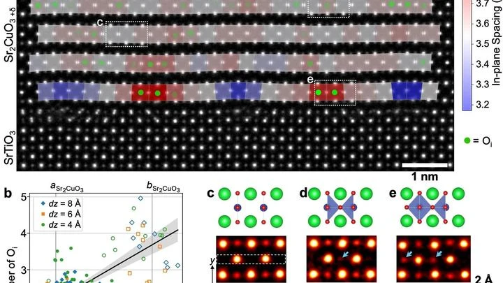
A superconductor is a material that conducts electricity without loss, like water flowing through a perfectly smooth pipe. In copper-based superconductors, tiny oxygen additions act like a volume knob: a bit more and the material starts superconducting; a bit less and it stops. But the oxygen atoms themselves are so small that seeing them directly was impossible.
A shipping container is a steel safe the size of a room, packed with goods. X-rays struggle to see through it: thick walls and dense cargo block the rays. Enter muons — particles born high in the sky that constantly rain down on Earth. They can pass through even lead, but they veer slightly off course when they encounter something heavy.
High-frequency sound focused on a single point acts like an invisible lens gathering sunlight. Inside a tumor, such a lens can raise the temperature to about 72°C in a second, like hot water from a tap, but inside living tissue. Meanwhile, surrounding healthy cells remain almost untouched.
A low-dose X-ray image looks like a view through a fogged-up window: outlines are there, but details drown in a hazy veil. Doctors need such images during radiation therapy — to aim the beam precisely at the tumor and spare healthy tissue. But saving on radiation comes at the cost of noise, streaks, and artifacts that obscure what matters.
Radiation therapy is a precision strike on a tumor, but every shot must be calibrated to within a millimeter and a fraction of a second. The problem is that existing dosimeters are either bulky, or they distort the beam themselves, or they require pauses for readout.
Tomographs don't produce ready-made pictures. They collect numerous 'shadows' — projections of innards from different angles or using various signals — and mathematically reconstruct a 3D picture from them.
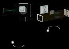
Each positron annihilation is like the birth of a pair of dancers, connected by an invisible thread of polarization. Two gamma quanta fly apart, but their states remain entangled: measuring one instantly determines the other — as if one partner senses every movement of the other, no matter the distance.
A radiologist stares at gray-white smudges on the screen — dozens of body slices where every suspicious spot must be found. Manual segmentation of one patient takes half an hour, and a large clinic sees hundreds of such patients. Fatigue and varying experience inevitably introduce variability: two specialists will outline the same lesion slightly differently.
In a small Petri dish sits a droplet of liquid the size of a pinhead. An ordinary scanner wouldn't even notice it: the signal is too weak. But bring a tiny magnetic field sensor close to the droplet, and the picture changes.
Ever since Paul Dirac theoretically predicted antimatter, physicists have been hunting for the tiniest differences between particles and their antiparticles.
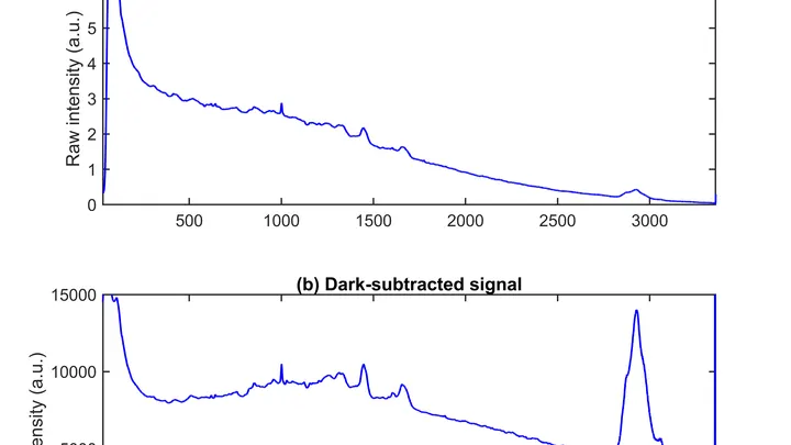
A drop of blood from a vein falls under three beams, and each one drags its own clues into the light. The infrared beam makes molecules vibrate — from that trembling, you learn how much sugar and fat is in the sample. A green laser scatters off proteins and lipids, shifting color — revealing the remodeling of cell membranes.
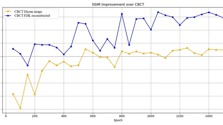
An X-ray tomograph is like a carpenter who examines a log from all sides to see where the void is inside. It takes dozens of flat shadow images, rotating the source and detector around the object. Then a 'reconstruction' program glues these shadows into a 3D model. The quickest reconstructor is the FBP algorithm (filtered back projection).
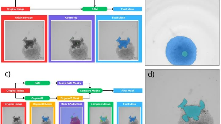
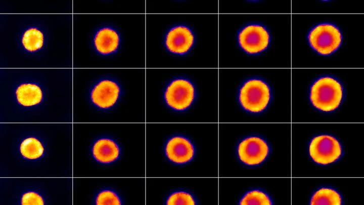
A biologist looks through a microscope at dozens of tiny cell spheres — spheroids. From these, miniature organ-like structures will later grow. The task is similar to a baker's job of checking whether the dough has risen evenly in each mold. Only here, instead of molds, there's a nutrient medium, and instead of dough, there are living cells.
A tongue tumor is like a stranger in a crowd. On the surface, they all look much alike, but beneath the surface hide very different personalities. Some tumors behave aggressively, others grow slowly. Doctors need to understand who they're dealing with before surgery.
Usually, this means taking a tissue sample — a biopsy.
A surgeon removes a breast tumor, but how do you know no cancer cells are left at the edge? Right now, the tissue goes to a lab, and the answer comes back days later. If the margin is not clean, a second surgery is needed. The new method promises to give the answer right in the operating room, while the patient is still on the table.
A tomograph is like a photographer who walks around an object and takes pictures from different sides. Usually, it snaps at regular intervals and uses the same amount of light for each shot. But if you need to shoot quickly and not waste extra film, you have to choose: which angles matter more and where to direct more light.
Two nearly identical locks are opened by the same pick. They look alike on the outside, but inside the mechanisms differ. Much the same can be said for the two main types of non-small cell lung cancer — adenocarcinoma and squamous cell carcinoma. Under a microscope, they can be confused, yet they require different treatments. That's where the quantum "magnifier" comes in.
Inside every cell, there are tiny power stations — mitochondria. They burn nutrients and store energy. Two helper molecules, NADH and FAD, oversee this process. Previously, they were detected by their faint glow, but it was dim, and signals from different molecules overlapped.
The new microscope works differently.
Inside every cell, there's a network of thin tubes — microtubules. They act as both scaffolding and rails: they hold the cell's shape and transport cargo. These tubes are built from the protein tubulin and naturally glow faintly under ultraviolet light. Scientists wondered: can we make this glow stronger or weaker on demand?
The key to control is the amino acid tryptophan.
A mole on your skin looks like a mosaic of colored pieces. Each piece is a small patch of the image where color and texture barely change. Such pieces are called superpixels. If you connect them with lines, you get a network that looks like a subway map: stations are the pieces, and lines are the connections between them. The weight of each line shows how strongly two pieces are related.
The camera that hunts for tumors works like a highly sensitive camera: it catches the faint glow from a substance that accumulates in diseased cells. In conventional machines, the light is born in a crystal — a solid piece that resembles glass. But a crystal doesn't always convey exactly where the light flashed, so the image comes out slightly blurry.
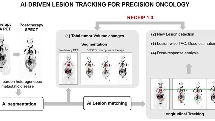
When treating prostate cancer, doctors often see not one tumor but many lesions in bones and lymph nodes. Each lesion can respond to therapy differently: one shrinks, another stays the same, a third grows. Previously, the disease was assessed as a whole, like judging the weather by the average temperature over a year.
An X-ray image is like a photo taken in the dark: the less light, the harder it is to see details. Doctors wanted to know how much you can dim the "light" in computed tomography (CT) and still not miss a disease. To do this, they took 50 pairs of chest images: one with a full radiation dose, the other with a low dose.
PET/CT is like a map where cancer cells light up as bright spots. But what if the map is blank and the disease is still hiding somewhere? In 132 men with recurrent prostate cancer, doctors found no glowing lesions, but they decided to study the texture of the scan itself, like an experienced tracker noticing bent grass where an animal has passed.
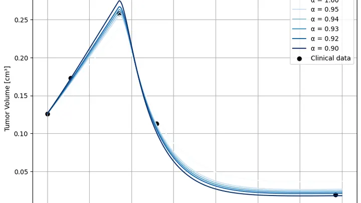
A tumor doesn't grow like a tree; it grows like a city: today's skyline is shaped by yesterday's construction and the fires of the day before. Standard tumor growth equations don't account for this: they only look at the current moment, as if the city were rebuilt from scratch every day. The authors added "memory" to the model: now the equation remembers what came before, and that changes the forecast.
A beam of carbon ions strikes the tumor precisely, like a surgeon who can only see the skin. Inside the body, the beam stops, and exactly where used to be a mystery. Now, helium is mixed into the same beam: it passes through the body like an X-ray, and its trail shows where the carbon went.
A lightning bolt strikes an anthill. Some ants die instantly, some freeze, and a few days later the survivors start rebuilding the tunnels. That's roughly how a tumor behaves after electroporation — a method where short electric pulses are passed through tissue to poke holes in cancer cell membranes and kill them.
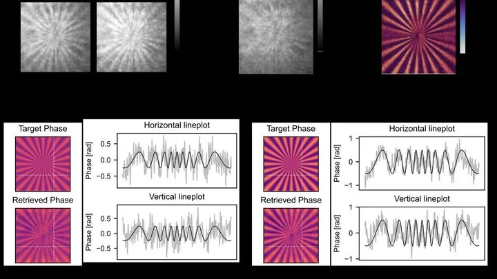
A glass marble in a glass of water is nearly invisible, but if you look through it at a checkered tablecloth, the lines behind it appear distorted. Transparent cells behave the same way: light passes through them but is slightly slowed and changes direction.
A firefighter with a thermal imager can see the source of a fire through smoke. But how do you see a radiation source that has no smell, no glow, and no heat? Gamma cameras solve this problem: they capture gamma rays emitted by radioactive materials and overlay their 'trace' onto a regular photograph. The result is an image showing that the source is, say, behind the far door.
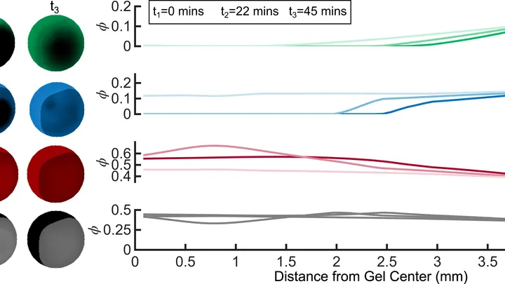
In winter, puddles freeze, and ice cracks the asphalt. Inside a living cell, freezing does something similar: ice crystals tear its membrane. To prevent this, the cell is soaked in a special solution, like antifreeze for a car. But precision is key: too little solution and ice still forms; too much and the solution itself poisons the cell.
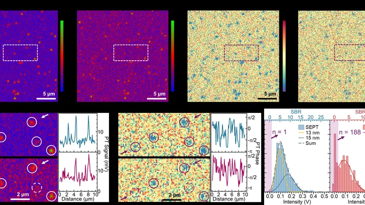
Light in a microscope doesn't arrive as a steady stream but like buckshot from a bag: sometimes more photons, sometimes fewer. This random scatter is the shot noise. It sets a limit: faint signals from tiny objects get lost in the ripple.
Scientists have found a way to calm this buckshot.
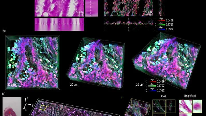
Light passing through a stained cell loses some of its strength, like a flashlight beam cutting through a glass of tea. The denser the color, the more light is absorbed. But until now, a microscope could only show a flat shadow of this process, not its volume.
Treating cancer with a proton beam is a bit like archery: you need to hit the bull's-eye without hitting anything else. Protons release their energy at the end of their path, so healthy tissues are largely spared. But to make the shot safe, every treatment plan is checked in advance — measuring the dose the tumor will get and what the surrounding organs will receive.
A drop of blood can reveal how a tumor in the lungs is behaving. Doctors have long noticed that when the disease retreats, some blood markers rise while others fall. But tying these changes into a single coherent picture has been elusive.
A glass of water and a glass of jelly look the same but behave differently. So it is with prostate tissue: on a standard slide under a microscope, you can see both the tumor glands and the supporting connective tissue around them. Previously, pathologists focused only on the glands, treating the support as mere background.