Microscope Sees Cells Like an Artist
A pathologist looks through the eyepiece and sees a pink-purple tissue section — the classic hematoxylin and eosin stain. The dyes bind to nuclei and cytoplasm, and the color tells the doctor about the disease. But preparing the slide itself takes hours: fixation, paraffin embedding, sectioning, staining.
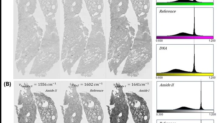
This new device skips the dye.