Near-infraredlight occupies the part of the electromagnetic spectrum just beyond visible red, with wavelengths roughly from 700 nanometers to a few micrometers. In biological tissue, this range is special: water and hemoglobin absorb visible light strongly, but near-infrared photons scatter less and encounter fewer absorbing molecules, allowing them to penetrate several centimeters deep. This 'optical window' makes near-infrared the go-to choice for non-invasive imaging and sensing inside living organisms, from pulse oximeters to brain activity monitors.
History
The optical window was recognized in the mid-20th century, with systematic studies by researchers like Britton Chance in the 1970s–80s who applied near-infraredspectroscopy to measure tissue oxygenation.
How it works
The key is that tissue components like water, lipids, and oxy- and deoxy-hemoglobin have low absorption coefficients in the near-infrared range, while scattering is relatively weak compared to visible light. Photons in this window undergo many random scattering events but still travel far enough to return to a detector, carrying information about absorption changes caused by blood flow or oxygen levels. In microscopy, near-infrared excitation also reduces photodamage and autofluorescence, enabling long-term observation of living cells.
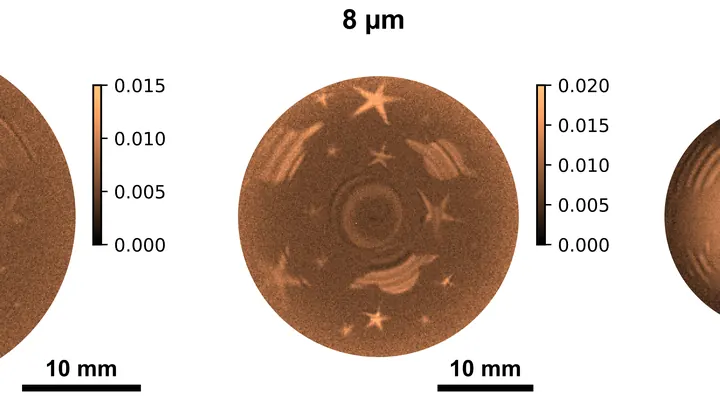
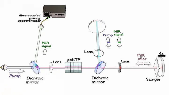
Light that never reaches the camera still leaves its mark on it. That's how the new imaging method works: you illuminate the object with one color, and the picture is assembled from another. Inside the crystal, light waves mix, and information about the invisible transfers to a visible beam.
In the experiment, they used a silver thiogallate crystal.
Light passing through a glass of murky water dims — some of it is absorbed. That's how many liquid-analysis instruments work: they measure how much light was lost and infer the composition from that. But if the liquid is heterogeneous, like soup with chunks, a single number isn't enough: you need to know where exactly more absorption happened.
A smartphone camera captures light with tiny sensors, but by themselves, they can't distinguish colors. So color filters are placed in front of them: red, green, blue, like stained glass in a window. Each sensor sees only its own color, and then software assembles the full picture from these pieces.
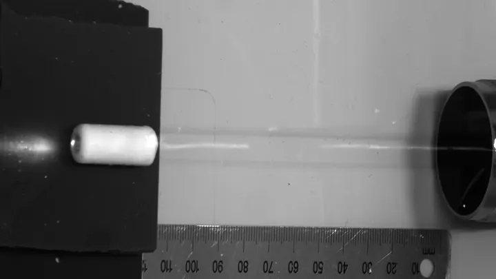
A synchrotron beam is like a jet of water from a hose: wide and awkward for fine work. To examine a tiny sample, you need a fine needle, not a hose. In the new setup, the needle is a glass capillary — a tube through which light glides, reflecting off the walls like a voice echoing down a long corridor.
When a star ventures too close to a supermassive black hole, a cosmic requiem plays out — a tidal disruption event. Thanks to photometry and spectroscopy, astronomers discern every instrument in this orchestra.
A typical laser is a cavity where light bounces between mirrors and gets amplified. But you can ditch the mirrors and make light circulate inside a tiny particle smaller than the light wave itself. That's how the new lasers work: nanoparticles of indium phosphide sitting on perfectly smooth gold.
The atmosphere of Titan, discovered by Christiaan Huygens, is opaque to ordinary telescopes. But spectral analysis finds 'windows' of transparency — narrow regions where the haze parts.
The James Webb Space Telescope peered into the era when the expansion of the Universe stretched the light of distant objects into the red, and found a scattering of strange sources — 'little red dots'. Their spectra broke the usual patterns: they are blue in the ultraviolet, but deep red in the optical, as if seen through a cherry veil.
The cosmic microwave background is the afterglow of the Big Bang, which we see at millimeter wavelengths. But it also has a long-wavelength tail extending into the radio range.
Neutron stars are the ultra-dense remnants of stars, where matter is compressed to the limit described by Subrahmanyan Chandrasekhar. Magnetars are a type of neutron star with a magnetic field a quadrillion times stronger than Earth's.
A high-speed camera capable of capturing the motion of electrons is an attosecond interferometer. It sends two laser pulses at a substance with a tiny delay. The first, ultraviolet, acts like a flash, knocking an electron out of the molecule. The second, infrared, like an invisible hand, gently nudges or slows this electron in flight.
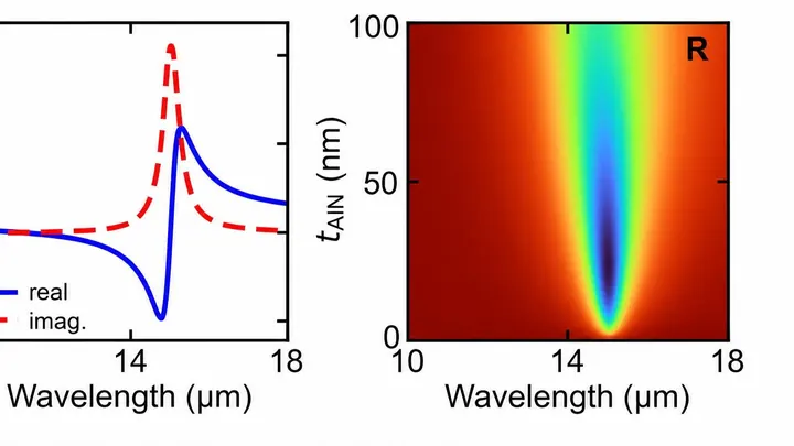
A film of aluminum nitride, as thin as a soap bubble, hangs above a mirror with a micrometer-sized air gap between them. When invisible infrared light with a wavelength of about 15 micrometers hits it, the film starts to vibrate in sync with the light, like a string that responds only to its own note.
Your finger is a living sensor: beneath the skin, arteries pulse in time with your heart. Traditional blood pressure cuffs squeeze your arm and give a single number, but this ring uses eight electrodes to pass a weak current through the tissue and map blood flow.
An ordinary flash drive stores bits in electron traps, but you can only read them through a separate wire and amplifier. In the new development, the same piece of the semimetal tellurium plays both the role of storage and the light bulb.
A sheet of graphene is a mesh of carbon atoms one atom thick. Even the slightest contamination shows up on it, like dust specks on eyeglass lenses. Ten years ago, researchers learned to hide graphene between layers of boron nitride, like placing a fragile sheet between two pieces of plastic wrap.
Infrared light is invisible to the eye, but it's everywhere: from TV remotes to the heat of the human body. Catching it requires special materials. Conventional detectors are bulky, rigid, and not easily tuned to specific wavelengths.
At night in a dark room, the human body emits a barely perceptible glow. This light is so faint that only highly sensitive detectors can catch it. Some researchers thought this glow from the head could tell us about brain activity — no scanners or wires needed.
But when put to the test, the story changed.
Microplastics, tiny fragments of synthetic polymers, are now found everywhere, from mountain glaciers to food. The traditional analytical approach, spectroscopy, which relies on light interacting with matter, has long required costly detectors to register invisible infrared radiation.
A green flashlight beam dives into a pool and reaches the bottom with barely any loss. Water is transparent to green light, which is why it's used for underwater communication. But to send video or sensor data, the light needs to be switched on and off very quickly — billions of times per second. Ordinary lasers are too slow and unstable for this.
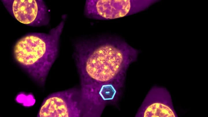
Light we can't see but feel as heat turns out to be the key to cellular life. A new type of microscope uses waves 2000–2500 nanometers long, three times longer than red light, and such waves barely scatter in tissue. Thanks to this, the device peers into a tumor spheroid like a flashlight through frosted glass, illuminating individual molecules.
A fat droplet inside a living cell is like a bobber in rough water. It doesn't move in a straight line but in jerks, sometimes pausing, sometimes speeding up. Previously, such movements could only be seen in a single plane, like watching the bobber from above without knowing its depth.
When treating epilepsy, doctors adjust the drug dose almost blindly. Too little, and seizures continue; too much, and side effects appear. Usually, a patient's blood is sent to a lab, and the result comes back a day or two later. Now, the same test can be done on a tiny silicon chip.
The sensor works like a highly sensitive nose.
An ordinary piece of semiconductor weakly changes the color of light passing through it. For decades, physicists worked around this limitation by placing the crystal in clever surroundings, but the material itself remained the same.
Now researchers have assembled a "sandwich" of alternating thin layers of gallium arsenide and aluminum gallium arsenide.
A distant galaxy birthing hundreds of stars should glow in oxygen. That's what astronomers thought: hot young stars heat the gas, which then emits a characteristic infrared glow at a wavelength of 63 microns. It's like steam rising from a boiling pot — a sure sign of vigorous activity.
Light in murky water behaves like a person in thick fog: every step is a collision, and the picture blurs. That's why a regular microscope can't see through skin or milk. Engineers found a workaround — look in the infrared range, where light collides less with particles.
A laser beam usually quivers slightly, like a plucked string. In optical clocks, that quiver turns into timing errors. Engineers swapped the shaky light for steady light, and rubidium vapor clocks started ticking five times more evenly.
A warm cup or a human hand emits invisible light — mid-infrared radiation. Every substance absorbs this light in its own way, leaving a unique 'fingerprint'. From this fingerprint, you can tell what an object is made of without touching it.
Previously, seeing this fingerprint required huge lasers that fired very short pulses. They were expensive and complex.
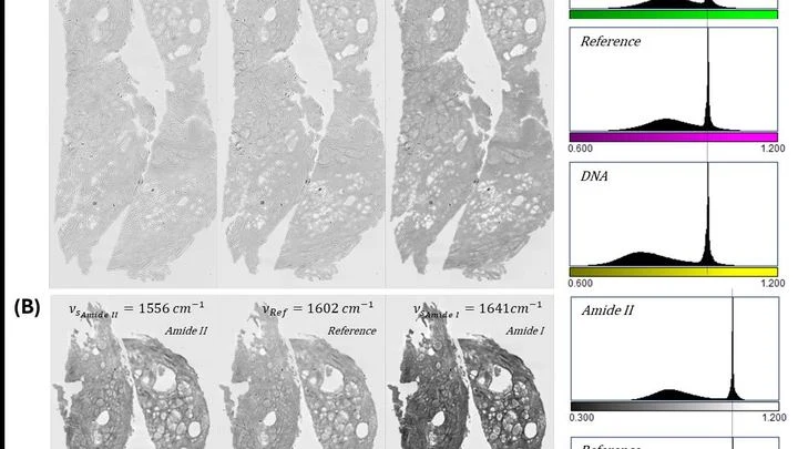
A pathologist looks through the eyepiece and sees a pink-purple tissue section — the classic hematoxylin and eosin stain. The dyes bind to nuclei and cytoplasm, and the color tells the doctor about the disease. But preparing the slide itself takes hours: fixation, paraffin embedding, sectioning, staining.
This new device skips the dye.
A light beam sent from Earth to a satellite scatters in the atmosphere, like a flashlight in fog. To keep the signal intact, you need short-wavelength light that spreads less. Such light is born in special crystals, but shrinking them to chip size is tough.
You can trap light in a cage smaller than a speck of dust. Photonic crystals are lattices that stop light from scattering and force it to run in circles. Previously, such structures were grown on a bulky substrate, making them too large for living cells.
There is a region of light that has long remained a blind spot: from 5 to 15 terahertz. It is not radio waves, nor infrared heat, but something in between, where molecules vibrate in particularly characteristic ways. Scientists from Germany have built a setup that finally peers into this 'new terahertz gap' and even beyond — past 25 terahertz.
The secret lies in a crystal with the long name BNA.
A quantum computer is like an orchestra that can only perform in complete silence. The slightest noise, and the delicate melody falls apart. For quantum devices, heat is that noise: even faint infrared radiation from nearby components heats up the qubits and erases their quantum states.
Light is a great tool for studying tissues, but it fades fast deep inside the body. Within a few millimeters, photons scatter so much that you can't make out anything. Scientists came up with a workaround: instead of shining light from outside, deliver a tiny light source right inside. The challenge is how to power that source without wires.
A rubidium atom is like a lock that opens only with one key: a particle of light of a strictly defined color. Even under the fiery rain of the sun, where myriads of colorful 'keys' fall, the lock remains silent. But when the right one strikes, the atom gets excited for an instant, and the detector registers a click.