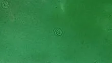
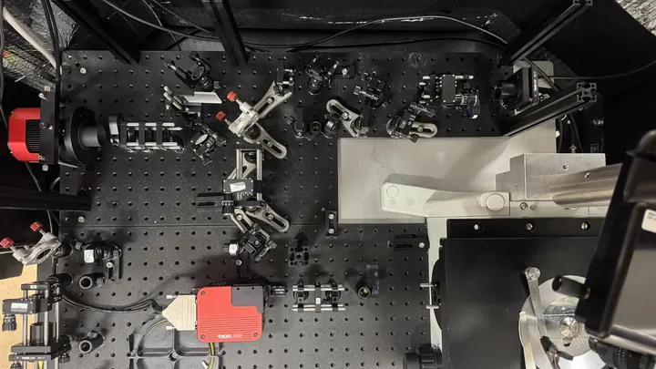
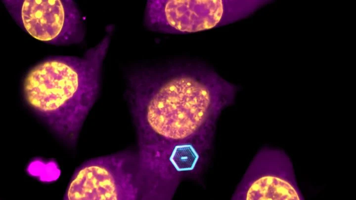
Live cell imaging is a set of optical techniques that let researchers watch biological processes unfold inside living cells, in real time, without killing the sample. Unlike traditional microscopy, which often requires fixing or staining cells, these methods use gentle illumination and fluorescent markers to track molecules, organelles, or even whole cells as they move, divide, and respond to their environment. It has become a cornerstone of cell biology, developmental biology, and drug discovery.
History
The field grew from the development of the green fluorescent protein (GFP) in the 1990s, which allowed researchers to tag proteins genetically and watch them in living cells.
How it works
The core idea is to make specific parts of a cell visible without disrupting it. This is usually done by attaching fluorescent proteins or dyes to molecules of interest, then illuminating the sample with light of one wavelength and detecting the emitted light at another. Advanced techniques like confocal microscopy, total internal reflection fluorescence (TIRF), and light-sheet microscopy reduce phototoxicity and improve resolution, allowing long-term observation of dynamic processes.
In practice
Live cell imaging is used to screen drug candidates, study cancer cell migration, and observe the dynamics of immune cells in real time.
Fun fact
Some live cell imaging techniques use so little light that a single cell can be observed continuously for days without significant damage.
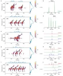
A conductor raises the baton, and the orchestra produces not just sound but a complex score: each note sounds at its own moment, at its own pitch, with the right volume. Light can also be a score, if you control its spectrum and timing. Previously, this required bulky setups that could not be reconfigured after assembly.
In a dark room, you can hold a glowing sphere without touching it: walls of light push it away but do not burn it. That is roughly how the trap built in Rio de Janeiro for yeast cells works. A conventional optical tweezer, that is, a focused laser beam, acts like tweezers made of light, but the light itself pierces the cell from side to side.
A thin section of eucalyptus lies under the microscope objective, and white light, passing through two polarizing filters, turns the wood into a mosaic of purple, yellow, and blue patches. These colors are not random: like letters on a map, they show the direction of the invisible cellulose threads inside the cell walls.
A navigator with a paper chart plots the route before setting sail. If the ocean changes—the current shifts, the wind picks up—the chart stays the same. The ship follows an outdated path, and the crew has to correct the course by hand.
A stenographer is recording a meeting, but halfway through the conversation she runs out of paper. She misses a few lines, then picks up where she left off. Later, she needs to reconstruct the full transcript without rewriting the entire conversation from scratch. That's exactly how the new method URA-SIM works for structured illumination microscopy (SIM).
Toss a handful of sand onto a table — the grains scatter equally in all directions. But if a magnet is hidden under the table and the sand is iron, the trajectories get trickier: an invisible structure overlays the chaos. That's roughly how molecular motion works inside a living cell.
Radiation therapy is a precision strike on a tumor, but every shot must be calibrated to within a millimeter and a fraction of a second. The problem is that existing dosimeters are either bulky, or they distort the beam themselves, or they require pauses for readout.
Data on gene activity usually looks like a long string of numbers, like a rainbow stripe (spectrum) with no visible connections. In the scVision model, these numbers are turned into a meaningful picture — not a list of ingredients, but a photo of a finished dish.
Dozens of copper coils in an MRI scanner pick up the faint response from tissues, like microphones trying to catch a whisper in a noisy hall. Each microphone drags a thick bundle of wires behind it. The wires press on each other, create electrical interference, heat up, and add risk for the patient.
A living cell constantly emits faint magnetic signals, as if whispering. Scientists eavesdrop on this whisper with two microscopic 'microphones'—magnetic field sensors.
Photographing a delicate spiderweb trembling in the wind: that's roughly what it takes to look into the blood vessels of a living brain. Ultrasound localization microscopy (ULM) turns ordinary ultrasound into a tool capable of seeing individual capillaries, but only if you get the settings just right.
In a benzene molecule, electrons buzz around the nuclei like a swarm of bees around a hive. But this swarm doesn't just hum: it pulses: every 856 attoseconds, the electron cloud completes a full oscillation cycle. The hive itself, the carbon nuclei, rocks more slowly, with a period of 36 femtoseconds.
A drop of ink in a glass of water spreads quickly and evenly. But if the water is squeezed between tightly packed spheres, the ink cloud crawls slowly and erratically: it dodges obstacles and gets stuck in narrow necks. Water molecules in living tissue move in much the same way.
A biologist looks through a microscope at dozens of tiny cell spheres — spheroids. From these, miniature organ-like structures will later grow. The task is similar to a baker's job of checking whether the dough has risen evenly in each mold. Only here, instead of molds, there's a nutrient medium, and instead of dough, there are living cells.
Your finger is a living sensor: beneath the skin, arteries pulse in time with your heart. Traditional blood pressure cuffs squeeze your arm and give a single number, but this ring uses eight electrodes to pass a weak current through the tissue and map blood flow.
Inside every cell, there's a network of thin tubes — microtubules. They act as both scaffolding and rails: they hold the cell's shape and transport cargo. These tubes are built from the protein tubulin and naturally glow faintly under ultraviolet light. Scientists wondered: can we make this glow stronger or weaker on demand?
The key to control is the amino acid tryptophan.
Biologists often tag cells with glowing molecules — it's like searching for a dim light in the smoke. The light quickly fades, and the smoke creates interference.
To avoid losing a fast soccer player in the frame, a drone doesn't need a bright beacon — recognizing the silhouette is enough. Similarly, a neural network now tracks microbes: the algorithm analyzes an ordinary microscope image and instantly finds a bacterium in a drop of water.
Strike a tuning fork, and it rings with a single pure note. But if the fork is trembling and people are chattering nearby, the sound won't be perfect. Same with molecules: each one absorbs light of a specific color, but due to its own vibrations and the jostling of neighbors, that color gets smeared out.
The authors of the paper figured out how to show this to students without complex formulas.
A typical accelerator is like an assembly line where electrons are the workpieces. To get the desired X-ray pulse, the workpiece is fine-tuned after it's already on the line: squeezed, filtered, corrected. In the new work, engineers decided to take a different approach — shaping the workpiece before it even hits the conveyor.
At LCLS-II, electrons are knocked out of a metal by a short laser pulse.
A fat droplet inside a living cell is like a bobber in rough water. It doesn't move in a straight line but in jerks, sometimes pausing, sometimes speeding up. Previously, such movements could only be seen in a single plane, like watching the bobber from above without knowing its depth.
A glass marble in a glass of water is nearly invisible, but if you look through it at a checkered tablecloth, the lines behind it appear distorted. Transparent cells behave the same way: light passes through them but is slightly slowed and changes direction.
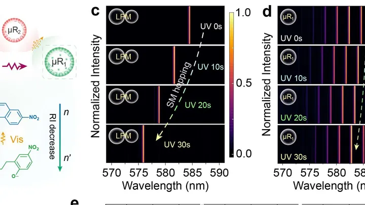
Two liquid droplets hanging side by side begin to sing in unison. One droplet is like a string tuned to a certain pitch, and together they form a laser that shines with a single, pure color. That's how a new type of sensor, a liquid photonic molecule, works.
The secret is that the droplets are slightly different in size.
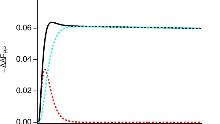
A heart cell in a Petri dish contracts, and at that moment, an electrical wave sweeps across its surface. Previously, to catch that wave, you had to poke the cell with an ultra-fine electrode needle. Now, you just look at it under a microscope.
The method is called electrochromic recording.
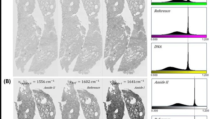
A pathologist looks through the eyepiece and sees a pink-purple tissue section — the classic hematoxylin and eosin stain. The dyes bind to nuclei and cytoplasm, and the color tells the doctor about the disease. But preparing the slide itself takes hours: fixation, paraffin embedding, sectioning, staining.
This new device skips the dye.
Cells breathe. They need oxygen, and they take it from the blood flowing through the tiniest vessels. If blood flow is weak or oxygen is scarce, cells start to suffocate. Doctors need to see this as early as possible. But how do you look inside living tissue without cutting it open?
The new device does it in two ways at once.
A tiny speck of silver is born in a laser beam and immediately starts to grow — faster than you can blink. An ordinary microscope can't keep up with that speed, so until now no one had seen exactly how a nanoparticle appears and gains size. Now scientists have a tool that captures this 'movie' frame by frame.
You can trap light in a cage smaller than a speck of dust. Photonic crystals are lattices that stop light from scattering and force it to run in circles. Previously, such structures were grown on a bulky substrate, making them too large for living cells.
A drop of blood is a bustling city in miniature. Thousands of different molecules scurry through it, each leaving its own trace. But peering directly into this city is hard: the streets are tangled, and there are so many inhabitants that counting them all individually is out of the question.
Scientists have found a way to listen to the whole city at once, without singling out its residents.
Inside every living cell, chemical transformations are constantly taking place. Some of them create unstable particles whose electrons behave like paired magnets. Their interaction resembles a dance: each movement of the pair responds to external magnetic fields, as if to a changing rhythm.
Living cells continuously exchange light signals — a billion times fainter than daylight. It's like a whisper picked up by ultra-sensitive instruments. A healthy cell whispers rhythmically, a cancerous one — haltingly, but with a long memory of previous flashes.