Light Pierces Milk: Lensless Microscope Sees Through Murky Media express
Light in murky water behaves like a person in thick fog: every step is a collision, and the picture blurs. That's why a regular microscope can't see through skin or milk. Engineers found a workaround — look in the infrared range, where light collides less with particles.
They assembled a lensless microscope — a device where a computer algorithm, not a glass lens, builds the image. A camera with a standard smartphone sensor captures light that passes through the sample, and software reconstructs the picture. Instead of visible light, they used near-infrared with wavelengths up to 1100 nanometers, like switching a flashlight from white light to an invisible but more penetrating beam.
Tests on mouse tissues showed: in the brain, structures are visible down to 250 micrometers; in the liver, down to 60. The camera sensor captures only 0.19% of photons at 1100 nm — light is barely registered, yet the algorithm still assembles a sharp image.
This work paves the way for cheap, compact devices for medical diagnostics that need no incisions or dyes. Light once deemed useless for such tasks now helps peer inside the living body.
Original abstract
as written by the authors arXiv:2602.14762
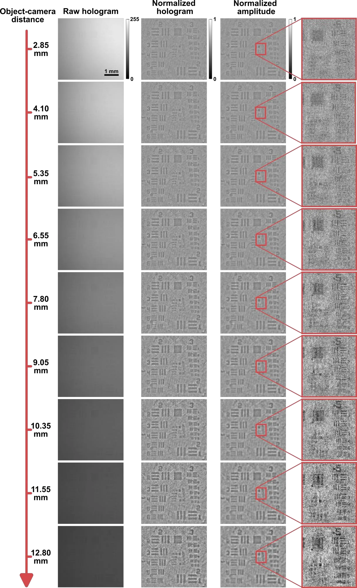
Lensless digital holographic microscopy (LDHM) relies on interference between an unscattered reference wave and a weakly scattered object wave - an assumption that rapidly fails in turbid samples under multiple scattering. To overcome this limitation, we present near-infrared LDHM (NIR-LDHM), a in-line holographic platform that operates up to the silicon cutoff (~1100 nm) using a conventional board-level CMOS sensor designed for visible (VIS) imaging. Using tissue-mimicking milk scattering layers and calibrated resolution targets, we quantify reconstruction performance versus wavelength, scattering strength, and sample-sensor distance. NIR-LDHM maintains resolvable features through scattering layers up to ~1.4 mm, whereas visible regime fails to resolve features below ~350 um. Importantly, despite a detector quantum efficiency of only 0.19% at 1100 nm, robust reconstructions are obtained under low-photon-budget conditions. We further identify advantageous mechanism by which increasing the sample-sensor distance from ~3 to 12 mm improves lateral resolution by twofold under strong scattering. Finally, we demonstrate wide-field, label-free amplitude-phase imaging of uncleared mouse tissues, resolving internal structure in brain and liver slices up to ~250 um and ~60 um, respectively. By extending lensless complex-field microscopy into strongly scattering regimes with minimal hardware changes, this work has relevance to computational imaging through complex media and biophotonics.
At 1100 nm, the camera sensor catches just one photon out of every five hundred, yet computational processing still yields a crisp image.
The idea of 'seeing through the murky' evokes sci-fi devices that scan the body without cutting — except here it's real technology based on an ordinary camera.