Non-invasive imaging refers to a broad class of techniques that allow us to see inside objects — living tissue, materials, or even the Earth — without physically cutting into them or inserting probes. Instead of a scalpel, these methods use various forms of energy, such as light, sound, magnetic fields, or microwaves, to probe the interior and reconstruct a picture from the signals that bounce back or pass through. This is the technology behind medical ultrasound, MRI, and X-ray CT scans, but it also extends to industrial inspection, geological surveying, and scientific microscopy.
History
The field began with Wilhelm Röntgen's discovery of X-rays in 1895, which allowed the first non-invasive look inside the human body. Later developments include ultrasound in the 1950s and MRI in the 1970s, pioneered by Paul Lauterbur and Peter Mansfield.
How it works
The core idea is to send a form of radiation or field into the object and measure how it interacts with the internal structure. For example, in MRI, a strong magnetic field aligns atomic nuclei, and radio waves are used to disturb them; as they relax back, they emit signals that vary by tissue type. In optical coherence tomography, near-infrared light is reflected off internal layers, and the time delay of the reflections reveals depth. The collected data is then processed by algorithms to create a two- or three-dimensional image, often without any physical contact beyond the surface.
In practice
Used routinely in hospitals for diagnosing fractures, tumors, and heart conditions, and in materials science for detecting flaws in aircraft parts or pipelines without damaging them.
Fun fact
Some non-invasive imaging techniques can now capture images at speeds fast enough to see the beating heart in real time, without any surgical intervention.
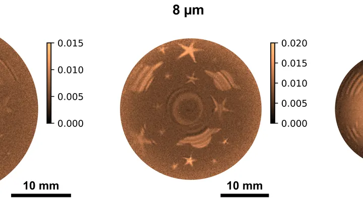
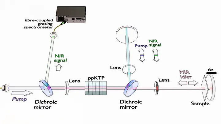
Light that never reaches the camera still leaves its mark on it. That's how the new imaging method works: you illuminate the object with one color, and the picture is assembled from another. Inside the crystal, light waves mix, and information about the invisible transfers to a visible beam.
In the experiment, they used a silver thiogallate crystal.
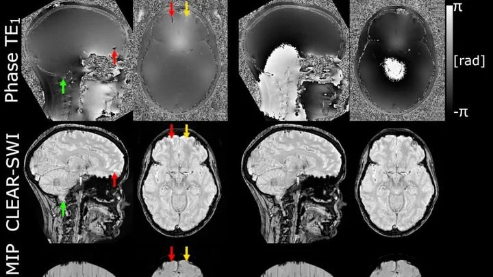
The powerful magnet of an MRI scanner induces weak electric currents in tissues. Their phase behaves like a compass needle trembling with every gust of wind — the slightest disturbance turns the map into a ripple, where instead of small vessels or iron deposits, all you see is noise.
A photographer shoots a runner with a long exposure and gets a blurry silhouette. That's roughly how a standard MRI scanner "sees" moving organs: the heart contracts, the lungs breathe, and the image smears. To avoid this, patients are asked to freeze and hold their breath. But for a child or someone short of breath, that's nearly impossible.
The method called GRASP tackles the problem differently.
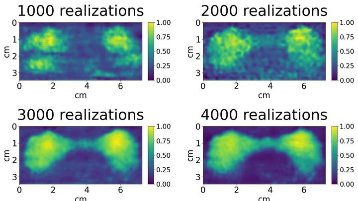
When you need to see inside the brain, you usually lie in a tight tube of an MRI scanner or get zapped with X-rays. Both methods are costly, bulky, and not always available.
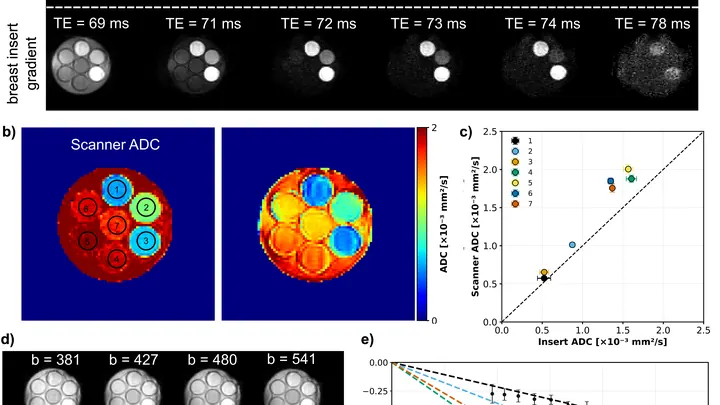
A flashlight spreads light in a broad beam, while a laser cuts a narrow path and picks out the finest details. In magnetic resonance imaging (MRI), the gradient, a rapid change in the magnetic field along the body, plays the role of that laser.
A drummer stays silent, but from the pauses you can tell where he didn’t hit. Similarly, physicists built an image by catching not light, but moments of absolute darkness. The picture emerged from silence.
In the experiment, they shone light from an ordinary lamp onto the object.
Microplastics, tiny fragments of synthetic polymers, are now found everywhere, from mountain glaciers to food. The traditional analytical approach, spectroscopy, which relies on light interacting with matter, has long required costly detectors to register invisible infrared radiation.
A finger is illuminated with a laser beam, and some of the light returns after traveling inside. That's how Raman spectroscopy works: molecules in bone and skin alter the color of the reflected beam differently, leaving their chemical fingerprints. But skin, muscle, and fat are noisy: their signal drowns out the bone's, like a crowd's chatter overpowering a single singer.
The boundary between two liquids is like an invisible skin: it's there, but you can't touch it with your finger. Scientists have found a way to measure its elasticity without touching it. They made a tiny probe oscillate above the surface, and between the probe and the interface lay a thin film of liquid.
A standard MRI machine is a huge magnet that you can't move, plus a powerful computer that turns signals from the body into a picture. In recent years, portable scanners with weaker fields have appeared: they are light, cheap, and can work in a field hospital or an ambulance.
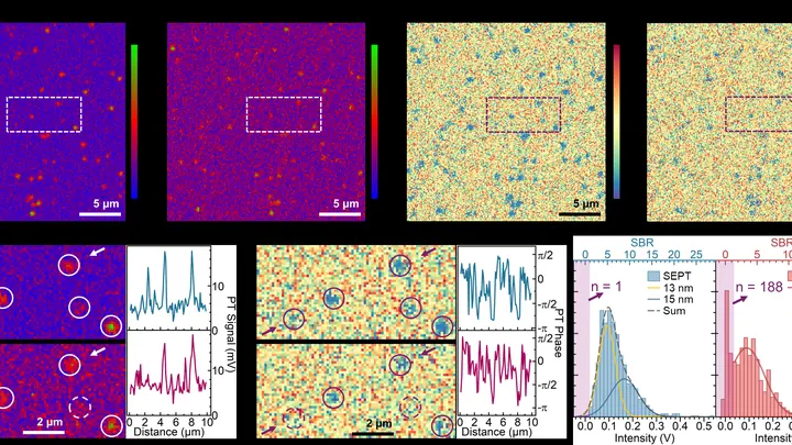
Light we can't see but feel as heat turns out to be the key to cellular life. A new type of microscope uses waves 2000–2500 nanometers long, three times longer than red light, and such waves barely scatter in tissue. Thanks to this, the device peers into a tumor spheroid like a flashlight through frosted glass, illuminating individual molecules.
A fat droplet inside a living cell is like a bobber in rough water. It doesn't move in a straight line but in jerks, sometimes pausing, sometimes speeding up. Previously, such movements could only be seen in a single plane, like watching the bobber from above without knowing its depth.
The heart doesn't contract all at once but as a wave: an electrical signal travels through the muscle, and behind it, like ripples spreading on water, a contraction rolls through. In arrhythmia, this wave goes awry: it stalls in one spot, loops in another, and the pump starts working in a disorganized way.
Light in murky water behaves like a person in thick fog: every step is a collision, and the picture blurs. That's why a regular microscope can't see through skin or milk. Engineers found a workaround — look in the infrared range, where light collides less with particles.
Radioactive tracers are like a courier with a beacon: you inject it into the body, and it highlights the target organ on the scan. But this courier comes with strict rules: you can't send it often, it quickly 'gets tired,' and it costs a lot. Doctors have long dreamed of a way to see an organ without radiation.
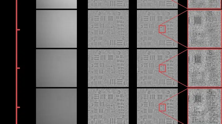
Light in a microscope doesn't arrive as a steady stream but like buckshot from a bag: sometimes more photons, sometimes fewer. This random scatter is the shot noise. It sets a limit: faint signals from tiny objects get lost in the ripple.
Scientists have found a way to calm this buckshot.
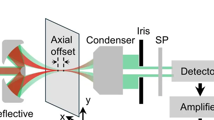
A warm cup or a human hand emits invisible light — mid-infrared radiation. Every substance absorbs this light in its own way, leaving a unique 'fingerprint'. From this fingerprint, you can tell what an object is made of without touching it.
Previously, seeing this fingerprint required huge lasers that fired very short pulses. They were expensive and complex.
The tip of a tunneling microscope hovers above a surface at a distance smaller than the diameter of an atom. Usually, it feels out the topography like a finger reading a page of Braille. Now, that same tip has learned to listen to how a membrane the size of a fingernail trembles.