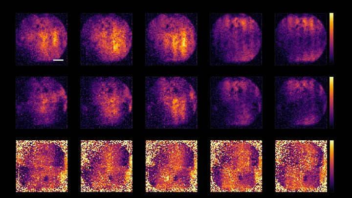
Noninvasive brain monitoring refers to techniques that assess brain function, structure, or pathology without penetrating the skull or inserting probes into brain tissue. These methods are essential for diagnosing and tracking conditions like brain tumors, Alzheimer's disease, and traumatic injuries, while minimizing patient risk and discomfort. Examples include optical spectroscopy, focused ultrasound, and advanced MRI protocols.
History
Noninvasive brain monitoring has roots in the early 20th century with electroencephalography (EEG), but modern optical and ultrasound methods have developed over the past few decades. The use of focused ultrasound for brain therapy has gained traction since the 2000s.
How it works
These techniques use external energy sources—such as light, sound waves, or magnetic fields—to interact with brain tissue and measure the response. For instance, optical spectroscopy shines near-infrared light through the skull and analyzes how it scatters and absorbs, revealing changes in blood oxygenation or tissue composition. Focused ultrasound can send sound waves through the skull to create images or even deliver therapeutic effects, while MRI uses magnetic gradients to map water molecules in the brain.
In practice
These techniques are used to monitor brain tumors during treatment, detect early signs of Alzheimer's disease, and guide therapies like focused ultrasound ablation without surgery.
Fun fact
A focused ultrasound beam can be shaped to pass through the skull and 'twist' inside the brain, allowing precise targeting of deep structures without damaging surrounding tissue.
In a fibrotic liver, as in a city after a long siege, neighborhoods appear where life has stalled: working cells are replaced by scar tissue, and hiding inside it are senescent macrophages. These cells have stopped dividing but have not retired. They release substances that push their neighbors to build even more scar and drive off immune patrols.
In a quiet room of a Budapest laboratory, a bracelet lies on the table, and instead of a strap, it has a thin conductive thread stretched in a zigzag. It doesn't heat up or glow, but when pressed against the wrist, it starts to listen. Each heartbeat sends a pressure wave through the radial artery, and the thread, deforming, changes its electrical resistance.
A drop of ink in a glass of water spreads in intricate swirls. Similarly, a quantum particle exchanging energy with its environment changes its state in a complex and entangled manner. Describing this dance between particle and environment has long baffled scientists.
Toss a handful of sand onto a table — the grains scatter equally in all directions. But if a magnet is hidden under the table and the sand is iron, the trajectories get trickier: an invisible structure overlays the chaos. That's roughly how molecular motion works inside a living cell.
Two scans of the same brain taken at different times are like photographs of the same house before and after renovation. To see what changed, you overlay them and look at the differences. But an MRI scanner is not a perfect camera: it adds its own distortions, as if the photo were taken through slightly warped glass.
During surgery, an anesthesiologist watches a screen where pressure and pulse curves run. But behind these lines hide several other quantities: how much the vessels constrict, how fast the blood flows, how elastic the artery walls are. Precise instruments for measuring them are only used in the most severe cases because it's risky — a needle in the artery, a catheter in the heart.
A flashlight shines through frosted glass: the rays bend, lose brightness, and take different paths. That's roughly how high-intensity ultrasound passes through the skull to the brain. For years, calculating this path required bulky computer models that 'chew' on data for hours.
A bell hidden in a thick box can be made to ring without opening the lid. Just send a sound wave through the walls, and the clapper will tremble. That's roughly how transcranial ultrasound works: sound passes through the skull bones and makes neurons deep in the brain 'ring'—generating electrical impulses.
A glucose meter that doesn't prick your finger but just looks at your wrist: that's the dream for millions of people with diabetes. It's the promise of algorithms that extract glucose levels from the ordinary pulse signal, the photoplethysmogram (the same one that measures heart rate in smartwatches).
Every time a child with cerebral palsy is about to move their leg, a distinctive electrical blip appears in the brain — a slow voltage shift. Scientists call it a movement-related potential. A brain-computer interface (a system that reads intentions from brain signals) can pick up this blip and turn it into a command for a training device.
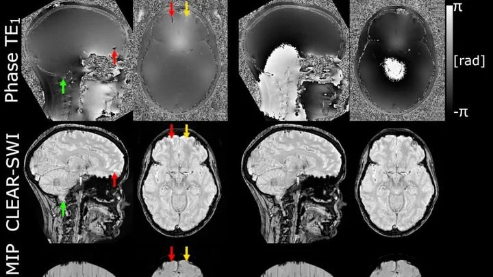
The powerful magnet of an MRI scanner induces weak electric currents in tissues. Their phase behaves like a compass needle trembling with every gust of wind — the slightest disturbance turns the map into a ripple, where instead of small vessels or iron deposits, all you see is noise.
Photographing a delicate spiderweb trembling in the wind: that's roughly what it takes to look into the blood vessels of a living brain. Ultrasound localization microscopy (ULM) turns ordinary ultrasound into a tool capable of seeing individual capillaries, but only if you get the settings just right.
A proofreader checks a manuscript before it goes to print. They don't know every possible mistake, but they know exactly what flawless text looks like. Spotting a strange symbol or a blurred word, they immediately circle it in red. That's roughly how the new program for checking medical images works.
Your arm lifts a cup from the shelf — a familiar motion you don't notice until muscles tire. Then the trajectory changes: the elbow shifts slightly, the hand trembles. A doctor usually catches these changes by hand, feeling the patient's muscles.
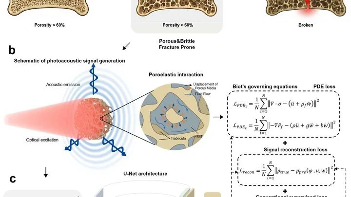
A laser beam strikes bone, and it responds with a nearly inaudible ultrasonic echo. Hidden in that sound is information about every microcrack and every fluid-filled pore. The trouble is that conventional computer programs don't understand this language: they see the signal but don't know the physics that produced it.
When you need to see inside the brain, you usually lie in a tight tube of an MRI scanner or get zapped with X-rays. Both methods are costly, bulky, and not always available.
If you check where an electron is every second, its quantum properties change. It's like a child playing hide-and-seek but constantly being called by name: they freeze and stop running. In the particle world, such a "caller" is a measurement. It prevents particles from freely forming a shared quantum state, or entanglement.
The brain is a fortress. It's protected by a dense layer of cells that only lets in what it needs. Drugs can't get through this barrier, which is why many brain diseases are so hard to treat.
Scientists have found a way to crack the fortress gates. They built a device that sends focused ultrasound into the brain. Tiny gas bubbles are injected into the blood.
Ultrasound penetrates bone like sunlight through frosted glass, but the image comes out blurry. The skull is uneven and dense, scattering sound waves, so hitting a precise spot in the brain is tricky. Conventional devices try to fix this with electronics, but the result is still far from perfect.
A surgeon removes a breast tumor, but how do you know no cancer cells are left at the edge? Right now, the tissue goes to a lab, and the answer comes back days later. If the margin is not clean, a second surgery is needed. The new method promises to give the answer right in the operating room, while the patient is still on the table.
The lungs are not two bags but millions of tiny sacs where air enters and blood flows close by. To see how they work, doctors sometimes use MRI without contrast: the scanner picks up weak signals from the tissues themselves. But these signals are like a radio in a thunderstorm — breathing is irregular, pulse jumps, and the image falls apart.
Your finger is a living sensor: beneath the skin, arteries pulse in time with your heart. Traditional blood pressure cuffs squeeze your arm and give a single number, but this ring uses eight electrodes to pass a weak current through the tissue and map blood flow.
Smartwatches measure your pulse with light: an LED shines through the skin, and a sensor picks up the reflected signal. By analyzing the fluctuations in this signal, you can estimate blood pressure, but accurate calculation requires a powerful neural network — usually running in the cloud, so data leaves the device.
The radioactive isotope terbium-149 acts like a tiny homing bomb: it latches onto cancer cells and detonates with alpha particles that destroy the tumor from within, barely touching healthy tissue. But this bomb has a quirk: it also glows with positrons, and that glow can be captured by a special camera.
Alzheimer's disease begins quietly: deep in the brain, two proteins, tau and amyloid-beta, start to accumulate. They behave like weed seeds: one clump that appears causes neighboring cells to produce similar clumps. Gradually, the proteins spread along nerve fibers, like water through the capillaries of a paper towel, and invade new areas.
A stroke hits suddenly, but before the strike, the brain sends quiet signals. Blood vessels stop smoothly regulating blood flow, like a thermostat that starts overheating or overcooling a room without reason. This imbalance is invisible on ordinary wearables, but it can be caught by listening to several body rhythms at once.
A finger is illuminated with a laser beam, and some of the light returns after traveling inside. That's how Raman spectroscopy works: molecules in bone and skin alter the color of the reflected beam differently, leaving their chemical fingerprints. But skin, muscle, and fat are noisy: their signal drowns out the bone's, like a crowd's chatter overpowering a single singer.
A beam of carbon ions strikes the tumor precisely, like a surgeon who can only see the skin. Inside the body, the beam stops, and exactly where used to be a mystery. Now, helium is mixed into the same beam: it passes through the body like an X-ray, and its trail shows where the carbon went.
A standard MRI machine is a huge magnet that you can't move, plus a powerful computer that turns signals from the body into a picture. In recent years, portable scanners with weaker fields have appeared: they are light, cheap, and can work in a field hospital or an ambulance.
The heart doesn't contract all at once but as a wave: an electrical signal travels through the muscle, and behind it, like ripples spreading on water, a contraction rolls through. In arrhythmia, this wave goes awry: it stalls in one spot, loops in another, and the pump starts working in a disorganized way.
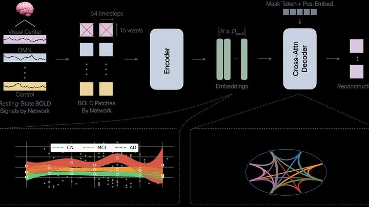
The brain isn't one big computer: it's more like an orchestra, where different groups of musicians play their parts. Neural networks for memory, attention, or emotions are constantly talking to each other. In Alzheimer's disease, this orchestra starts to go off-key: some groups fall silent, others play out of sync.
An electrocardiogram is like a long strip of paper on which the heart leaves its signature. A doctor studies it to catch rhythm disturbances, but when the recording lasts for days, the strip stretches for miles. The eye tires, attention wanders, and rare but dangerous irregularities can slip by.
AI-HEART works like a tireless assistant that scans the entire strip without a break.
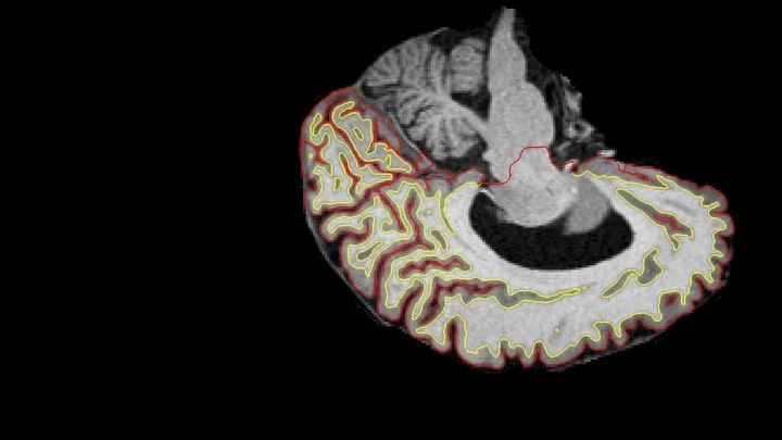
Every human brain is covered with folds, much like a fingerprint: each with its own unique pattern. In dementia, this pattern changes, but for two similar diseases, Alzheimer's and dementia with Lewy bodies, the changes are nearly indistinguishable to the eye. Doctors need a reliable way to separate them, because the treatments differ.
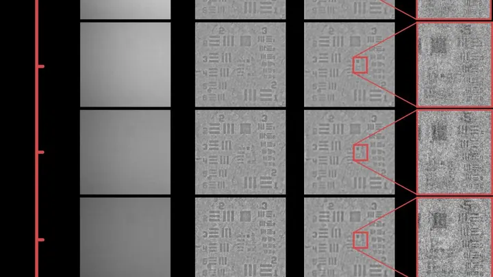
Light in murky water behaves like a person in thick fog: every step is a collision, and the picture blurs. That's why a regular microscope can't see through skin or milk. Engineers found a workaround — look in the infrared range, where light collides less with particles.
Your back hurts, and the doctor asks you to bend forward and backward to see how your vertebrae move. Usually, this means a series of X-rays: they show motion, but only in a plane, like a shadow on a wall. And volumetric methods like CT scans can't keep up with movement.
Engineers proposed a different approach: a robot sweeps an ultrasound probe over your back while you bend.
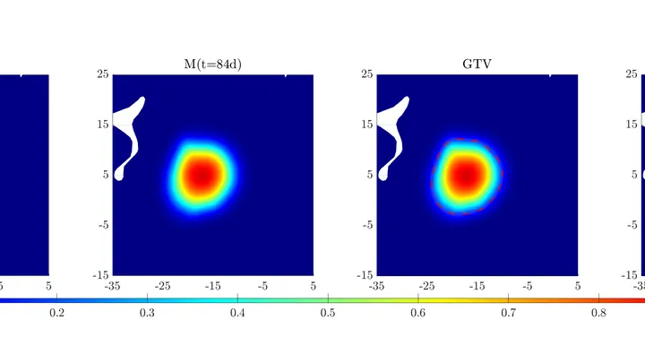
A brain tumor is like a traveler trudging through a dense forest: trees and bushes are the different types of brain tissue, some easier to traverse than others. Glioma cells don't move in a straight line; they constantly loop around, bumping into obstacles and going around them.
A tiny defect in diamond is like a microscopic ear, able to pick up the magnetic 'breath' of the heart. Such sensors work at room temperature and are no bigger than a grain of sand.
Treating cancer with a proton beam is a bit like archery: you need to hit the bull's-eye without hitting anything else. Protons release their energy at the end of their path, so healthy tissues are largely spared. But to make the shot safe, every treatment plan is checked in advance — measuring the dose the tumor will get and what the surrounding organs will receive.
A detective is looking for a criminal in a file of mugshots, but the portraits are mixed up. Usually, they expect a man with a gray beard, but the culprit is young and clean-shaven. Similarly, doctors often fail to recognize Alzheimer's disease in those who show unusual symptoms — not memory loss, but problems with vision or speech.