Movies for the Nanoworld: Watching Atoms in Motion express
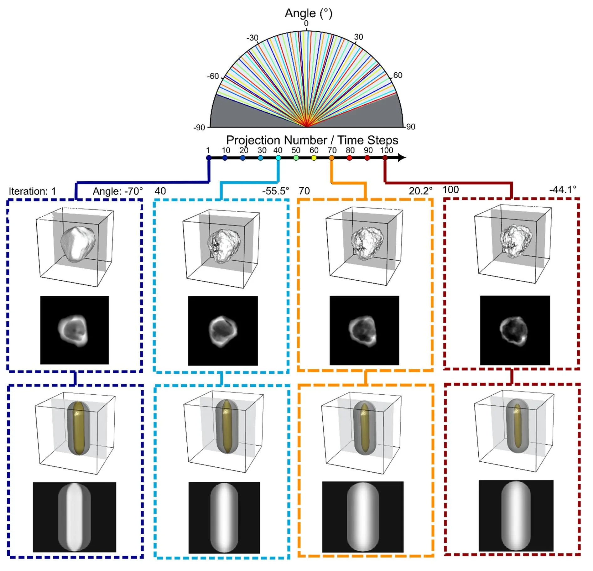
A tiny gold star melts under the beam of an electron microscope, its points slowly rounding off. In the past, such a process could only be seen in snapshots taken with long pauses, and the probing beam itself would alter the sample. Now, researchers shoot continuous footage: the sample tilts smoothly, and an algorithm assembles a three-dimensional picture from that stream for any point in time.
The secret is that the tilt never stops, and the computer learns from the data itself, without any hints. It understands how the particle's shape changes and fills in the gaps between frames. The electron dose is lower than in older approaches, so fragile structures, like silver nanoparticles with a gold shell, survive until the end of the recording.
The method was tested on two scenarios: how gold nanostars lose their sharp points when heated, and how gold and silver atoms mix inside a nanocube. In both cases, the evolution could be followed in 3D, not just on the surface. It's like watching not the shadow of a dancer, but the dance itself from all sides.
Now materials scientists can watch catalysts, batteries, or drug nanoparticles change right as they work, and do so gently, without worrying that the tool itself will ruin the experiment.
Original abstract
as written by the authors arXiv:2603.29462
Direct observation of nanoscale transformations in three dimensions (3D) is essential for understanding materials evolution under operating conditions, yet dynamic electron tomography remains limited by slow tilt series acquisition and by reconstruction methods that assume static specimens. These constraints prevent continuous 3D imaging of evolving structures and require electron doses that can alter the specimens and their dynamics. Here, we introduce a framework for dynamic electron tomography that combines continuous tilting with a self-supervised deep-learning reconstruction strategy. Our approach incorporates the temporal aspect into the electron tomography reconstruction process to recover 3D volumes at arbitrary time points from a single tilt series. We validate the method using simulations and demonstrate its merit in experimental studies of heat-induced transformations, including morphological evolution of Au nanostars and alloying in Au@Ag nanocubes. Our results establish a practical framework for dynamic, dose-efficient electron tomography, enabling in situ 3D investigation of nanomaterial transformations as well as the characterization of beam-sensitive structures.
A gold nanostar smaller than a hundred nanometers loses its sharp points when heated and becomes droplet-like: this process is now visible in 3D and in real time.
This technique is reminiscent of cameras from science fiction films that capture holograms of moving objects, but here it all happens in the world of atoms.