Scintillator in a new package: light becomes four times brighter express
A crystal the size of a matchbox was placed under the beam of a clinical accelerator. Half of its surface was covered with a pattern of nanoscale bumps — tiny, smaller than the wavelength of light. The other half was left smooth. The difference was striking: the coated part produced a signal 4.1 times brighter.
In radiation therapy, it is crucial to know exactly how much energy the tumor and healthy tissues receive. Scintillators convert radiation into visible light, but some of it is lost inside the crystal. The nanostructure acts like a tiny lens: it helps light escape rather than reflect back. The researchers verified that the coating did not distort the linearity of the response and did not introduce dose-rate dependence. The crystal remained an honest dosimeter.
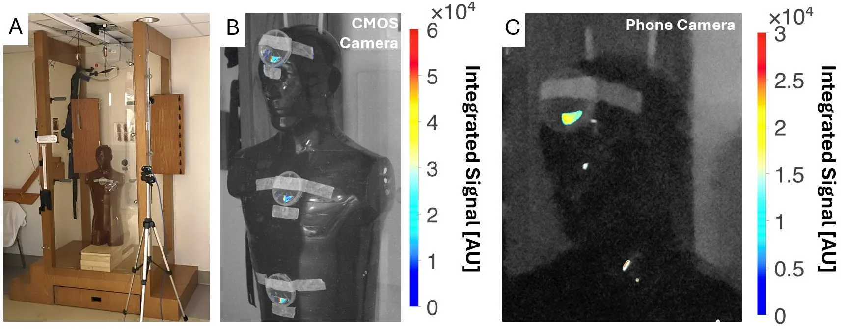
The final test was performed on a phantom in a room for total body irradiation. Light from the nanophotonic half was clearly visible both on a scientific CMOS camera and on a regular smartphone — even with room lights on. The smooth half was silent. The sensitivity was enough to see the signal without sophisticated equipment.
Original abstract
as written by the authors arXiv:2606.16007
Purpose: Recent advances in metasurface photonics and manufacturing have enabled a nanophotonic surface coating to be applied to conventional scintillators, which has been shown to significantly improve light yield. However, the dosimetric properties of such coatings has not been established. We performed the first dosimetric characterization of a nanophotonic scintillator and explored clinical application to real-time in-vivo total body irradiation (TBI) dosimetry. Methods: A 4.5x1.5 cm cerium-doped yttrium aluminum garnet (YAG:Ce) scintillator, half patterned with the nanophotonic structure, half left unpatterned, enabled direct comparison between conventional and nanophotonic surfaces. The scintillator was placed in a 3D-printed light-tight box with an off-axis CMOS camera and irradiated with a clinical linear accelerator. Absolute dosimetry was done with dose-calibrated radiochromic film. For TBI, the scintillator was positioned on an anthropomorphic phantom in a TBI booth, with signal measured by both CMOS and consumer-grade phone cameras under different room lighting conditions. Results: The nanophotonic scintillator showed a 4.1x increase in signal and a 3.7x increase in contrast-to-noise ratio versus the conventional scintillator. Both scintillators exhibited dose-rate independence and linear dose response, with modest energy dependence. For TBI, the nanophotonic scintillator produced a clearly detectable signal with both CMOS and phone cameras, whereas the conventional scintillator signal was undetectable. Conclusions: Nanophotonic structures significantly enhance light output of conventional scintillators without impacting their dosimetric properties. Nanophotonic scintillators may enable real-time in-vivo TBI dosimetry. Future work should investigate this technology for improved dosimetry equipment and X-ray imaging detectors.
The metasurface-based coating works like the anti-reflective coating on eyeglasses: it suppresses unwanted reflections and lets more light through.