How droplets inside cells shape membranes express
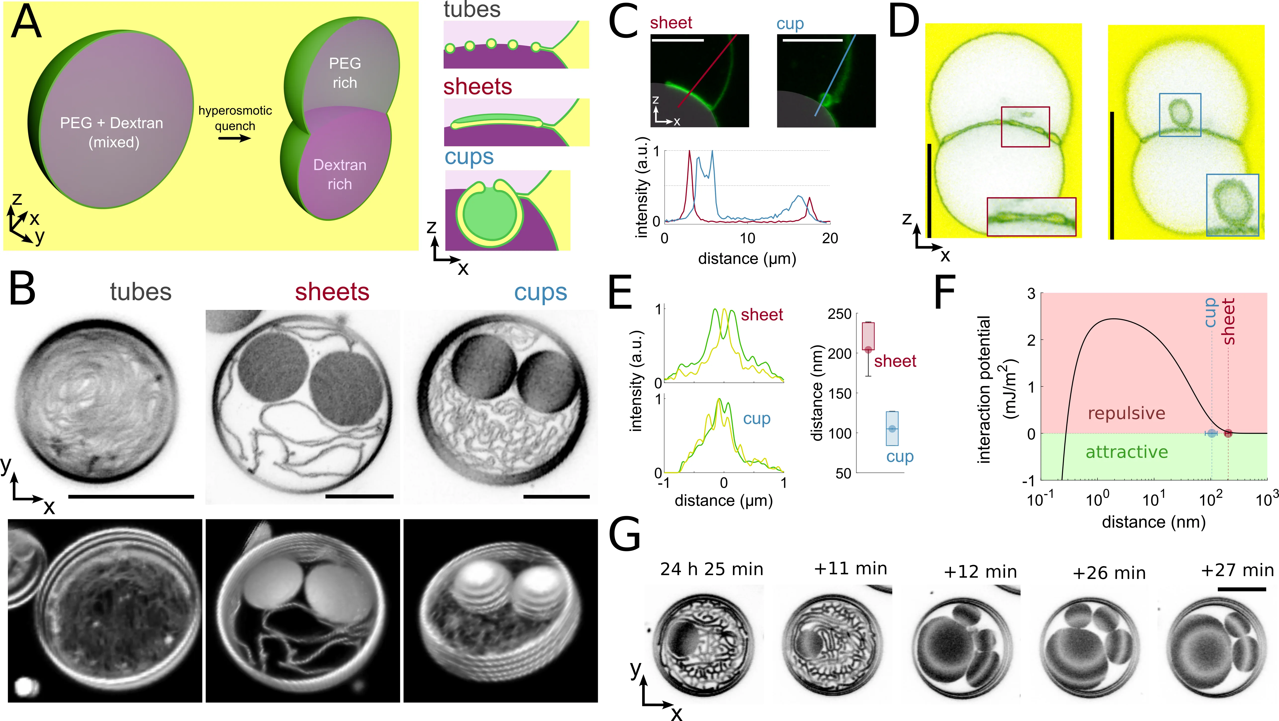
Inside a living cell, droplets float that don't mix with the surrounding fluid, like oil in water. These droplets aren't just passive clumps; they actively press on membranes and make them bend. Scientists observed this process in living plant cells, reproduced it in a test tube, and simulated it on a computer.
It turns out that the membrane's shape depends on the tension at the droplet's surface. If the tension is high, the membrane flattens into a sheet. If the tension is lower, the membrane rolls into a tube or a cup. The transition between these shapes isn't always reversible: to go back from a cup to a tube, you need to overcome an energy barrier, and the membrane 'remembers' its previous shape.
This mechanism explains how 'onions' form inside vacuoles in plant cells — large fluid-filled bubbles. The vacuole membrane invaginates under the influence of droplets and forms cup-shaped structures. By controlling the surface properties of the droplets, the cell can mold membranes into desired shapes at the right time.
Original abstract
as written by the authors arXiv:2603.15904
Phase-separated biomolecular condensates with liquid-like properties play a key role in the organization and compartmentalization of the intracellular environment. Condensate-mediated capillary forces acting on membranes drive physiologically important reshaping of membrane-bound organelles, such as vacuoles and autophagosomes. Here, we explore condensate-mediated membrane shape transformations. We employ {\textit{in planta}} live-cell imaging, an \textit{in vitro} reconstitution system with tunable interfacial tension, and computer simulations of an elastic membrane model to describe three morphologies of membrane structures localized at condensate interfaces: tubes, sheets, and cups. We find that the forces associated with high interfacial tension drive the formation of stable sheets, while tubes and cups prevail at lower interfacial tension. We calculate the free energies of each membrane shape and identify the energy barriers that govern the transitions between the shapes. With this approach, we find that shape transformations depend on the history of the interfacial membrane and exhibit a tube-to-cup hysteresis. These findings indicate that temporal control of condensate surface properties can mediate the morphogenesis of cup-like structures in cells, such as the formation of "bulbs" within plant vacuoles. Our results further generalize how the interplay of condensates and membranes contributes to intracellular organization.
In plant cells, vacuoles can occupy up to 90% of the cell's volume, and the droplets inside them help form complex internal structures.