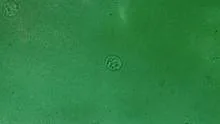
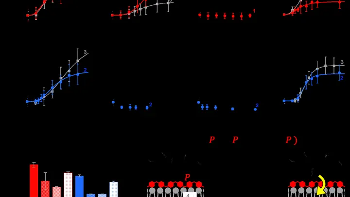
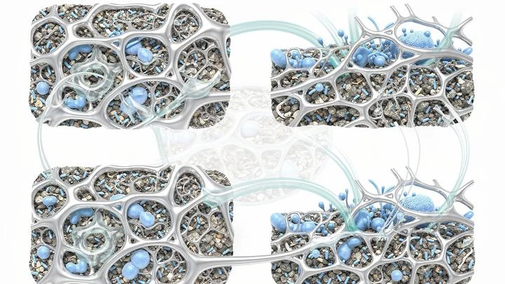
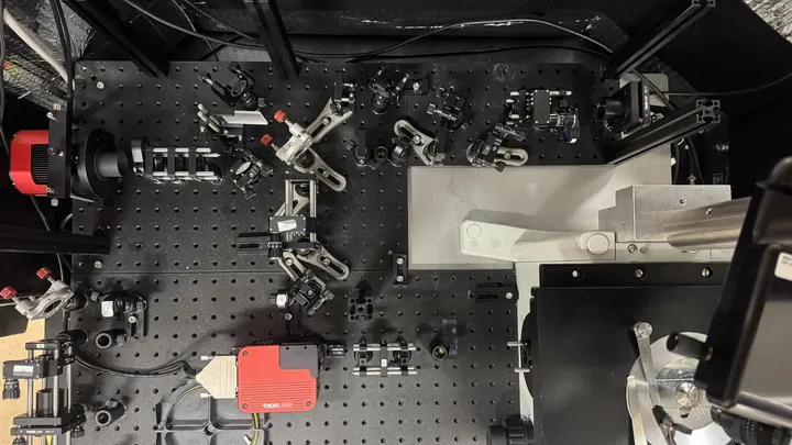
Living cells are the fundamental units of life, capable of metabolism, growth, reproduction, and response to stimuli. In physics research, they are studied as complex systems that exhibit mechanical, electrical, and optical properties. Cells are often sensitive to light damage, which is a critical consideration in microscopy and phototherapy. Understanding how cells respond to radiation and light is essential for medical diagnostics, cancer treatment, and biophotonics.
History
The discovery of cells is attributed to Robert Hooke in 1665, who observed cork under a microscope. The cell theory was formalized by Matthias Schleiden and Theodor Schwann in 1839. The study of cell light sensitivity has evolved with advances in microscopy and photobiology.
How it works
Cells are composed of a membrane, cytoplasm, organelles, and genetic material. They maintain homeostasis through biochemical reactions and can communicate via chemical signals and even light emission (biophotons). When exposed to light, cells may absorb photons, leading to photochemical reactions that can cause damage (e.g., DNA breaks) or trigger protective responses. The sensitivity depends on the wavelength, intensity, and duration of exposure, as well as the cell type and its repair mechanisms.
In practice
Living cells are used in medical diagnostics (e.g., blood tests), drug development, tissue engineering, and in research on photosynthesis and biophotonics for non-invasive health monitoring.
Fun fact
Some single-celled algae can act as living lenses, focusing light onto their chloroplasts to optimize photosynthesis, a phenomenon that has inspired optical engineering.
Every fall, Issaquah Creek in Washington State turns into a maternity ward for thousands of salmon. The fish swim in, spawn, and die, and the water carries away their molecular traces: fragments of DNA, short-lived RNA, chemical marks on the genetic text. From September through November 2024, researchers collected a liter of water every week and ran it through a filter to catch those traces.
In a dark room, you can hold a glowing sphere without touching it: walls of light push it away but do not burn it. That is roughly how the trap built in Rio de Janeiro for yeast cells works. A conventional optical tweezer, that is, a focused laser beam, acts like tweezers made of light, but the light itself pierces the cell from side to side.
A thin section of eucalyptus lies under the microscope objective, and white light, passing through two polarizing filters, turns the wood into a mosaic of purple, yellow, and blue patches. These colors are not random: like letters on a map, they show the direction of the invisible cellulose threads inside the cell walls.
The liver is an orchestra in which every cell plays its part from a shared score. Males and females have different scores: in males, the genes responsible for detoxifying foreign substances and steroid hormones sound louder, while in females, the genes for fat metabolism and bile acids do.
Life begins with a bubble. In a warm puddle on ancient Earth, fat molecules could gather into tiny spheres — prototypes of cells. Inside such a sphere, a polymer happened to be present that accelerated the uptake of new fats from the surrounding water. The bubble grew, divided, and passed its composition to offspring.
Toss a handful of sand onto a table — the grains scatter equally in all directions. But if a magnet is hidden under the table and the sand is iron, the trajectories get trickier: an invisible structure overlays the chaos. That's roughly how molecular motion works inside a living cell.
Imagine a packed subway car at rush hour. Passengers don't just stand there: they breathe, release moisture, squeeze toward exits, and sometimes get stuck in doors, blocking movement. Now imagine these passengers can remodel the car's walls, add handrails, or turn the floor into gel.
In embryonic development, a cell resembles a musician in a vast orchestra: it must play its part, following a conductor — the gradient of signaling molecules. But the conductor is unreliable: morphogen concentrations constantly fluctuate. How then does a cellist not veer into a violin melody?
A living cell constantly emits faint magnetic signals, as if whispering. Scientists eavesdrop on this whisper with two microscopic 'microphones'—magnetic field sensors.
In 1944, Erwin Schrödinger asked the question "What is life?", foreshadowing quantum explanations of biology. Almost a century later, physicists return to this puzzle, armed with the apparatus of quantum field theory.
Inside every cell, construction never stops: molecules constantly cluster together and drift apart. Sometimes they form dense droplets, like oil in water, but without a membrane. These droplets act as temporary storage: they hold proteins, gene copies, or DNA repair tools.
Life is a constant dance between order and chaos. Back in the mid-20th century, Erwin Schrödinger noted that living things feed not so much on matter as on "negative entropy"—the ability to maintain their own order by increasing disorder around them.
Billions of tiny motors hum inside every living creature. Cells ceaselessly pump substances, burn sugar, and build new proteins, and all this invisible motion warms us from within. To maintain order, life must dissipate heat into the environment, like a stove that never goes out.
The power of this internal heating is almost independent of size.
A tightrope walker inches across a chasm. As long as they keep their balance and avoid sudden moves, they're safe. But one overcorrection, and a fall is inevitable. Life works much the same way at every level — from proteins inside a cell to entire ecosystems.
The Standard Model is a set of rules for all known particles. But it says nothing about dark matter, which makes up a quarter of the Universe. To find it, you don't need to build city-sized colliders. Radioactive molecules act like sniffer dogs: their incredible 'scent' picks up the faintest traces of unknown forces that slip past giant machines.
In the freshwater lakes, cyanobacteria Microcystis often cause toxic blooms. They gather into colonies — elastic little spheres, like dense jelly. The cells are glued together by a special substance based on carbon, acting as a powerful natural adhesive. Scientists tested these "jelly balls" for the first time and found their strength astonishing.
The tiny green alga Chlamydomonas spends its life rowing with two flagella toward light, like a swimmer heading for shore. It senses light with a photoreceptor protein, and behind it lies a red spot — the 'eyespot'—which casts a shadow and helps the cell figure out where the light is coming from.
When you cut your finger, skin cells start working in concert: some divide, others crawl toward the wound, and still others pause. But exactly how a cell decides what to do next depends on its internal "schedule"—the cell cycle.
4 billion years ago, the young Earth was mercilessly bombarded by meteorites, leaving behind impact craters. Rains filled them with water, forming shallow ponds where lipid bubbles accumulated. These bubbles, like soap bubbles but with a double-layered lipid membrane (just like modern cells), formed spontaneously when water mixed with fat-like substances.
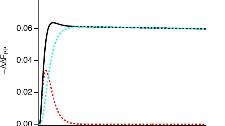
A heart cell in a Petri dish contracts, and at that moment, an electrical wave sweeps across its surface. Previously, to catch that wave, you had to poke the cell with an ultra-fine electrode needle. Now, you just look at it under a microscope.
The method is called electrochromic recording.
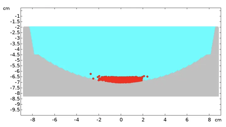
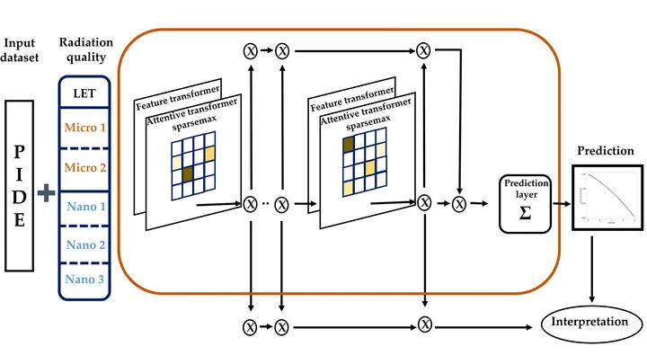
A cell hit by a proton beam is like a city after a pinpoint bombing: some blocks are leveled, others just have broken windows. Whether the city survives depends on which buildings are hit and how the strikes are distributed.
Inside every living cell, chemical transformations are constantly taking place. Some of them create unstable particles whose electrons behave like paired magnets. Their interaction resembles a dance: each movement of the pair responds to external magnetic fields, as if to a changing rhythm.
Living cells continuously exchange light signals — a billion times fainter than daylight. It's like a whisper picked up by ultra-sensitive instruments. A healthy cell whispers rhythmically, a cancerous one — haltingly, but with a long memory of previous flashes.