The Shadow of Cancer on a Clean Scan express
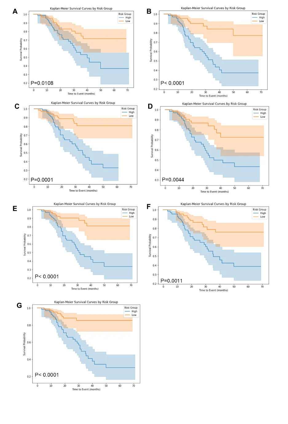
PET/CT is like a map where cancer cells light up as bright spots. But what if the map is blank and the disease is still hiding somewhere? In 132 men with recurrent prostate cancer, doctors found no glowing lesions, but they decided to study the texture of the scan itself, like an experienced tracker noticing bent grass where an animal has passed.
It turned out that even without bright spots, the tissue pattern in organs where cancer tends to spread (bones, lymph nodes) differs in those whose disease later progressed. Computer analysis of these subtle patterns, radiomics, combined with standard tests gave a more accurate prognosis than old methods: the chance of correctly predicting disease progression rose from 65 to 74 percent.
Importantly, the method remained accurate when tested on scans from another hospital. So it's not about the quirks of one machine or one doctor. This approach could help identify earlier which patients need closer monitoring or treatment, even when the standard scan looks reassuring.
Original abstract
as written by the authors arXiv:2603.19368
In patients with biochemical recurrence of prostate cancer and negative PSMA PET/CT, radiomics features extracted from recurrence-prone organs can predict clinical progression and progression-free survival. In a cohort of 132 patients, combining PET/CT radiomics with clinical variables significantly improved prognostic performance (C-index 0.74 vs. 0.65). Model performance was influenced by reader diagnostic certainty and remained robust on external validation. These findings suggest that radiomics from visually negative scans capture subclinical disease and provide added value for risk stratification.
The term 'radiomics' only appeared in 2012, but already algorithms are finding signs of disease on scans that even experienced radiologists can't see.