The PI-RADS (Prostate Imaging Reporting and Data System) is a standardized method for assessing the probability of clinically significant prostate cancer based on multiparametric MRI. Each detected lesion in the transition or peripheral zone receives a score on a 5-point scale. The assessment is based on analysis of T2-weighted imaging (dominant sequence for the transition zone), diffusion-weighted imaging (dominant for the peripheral zone), and dynamic contrast enhancement (which plays a supporting role). The rules for assigning the final score depend on the zone and the presence of certain features: border clarity, size, signal intensity. PI-RADS 3 indicates an uncertain result, PI-RADS 4 and 5 indicate the need for biopsy.
History
The need for unification arose in the early 2000s with the increasing use of mpMRI. The first PI-RADS version was released by ESUR in 2012 but was imperfect due to convoluted algorithms. In 2015, together with the American College of Radiology (ACR) and the AdMeTech Foundation, version 2 was presented, simplifying the rules and separating the approach for the transition and peripheral zones. In 2019, an interdisciplinary expert group released PI-RADS v2.1, refining criteria for the transition zone (for example, removing the 2+1 score, relaxing requirements for nodule shape), which increased specificity. PI-RADS is recognized as an international standard, and its validation continues in different populations.
How it works
After obtaining mpMRI images, the radiologist evaluates each finding sequentially: 1. Determine the zone (peripheral or transition) using T2 anatomy. 2. Peripheral zone: primary DWI assessment. DWI score: 1 — no changes, 2 — linear/wedge-shaped vague hypointensity, 3 — focal moderate hypointensity (indistinct, small), 4 — distinctly hypointense lesion <1.5 cm, 5 — distinctly hypointense lesion ≥1.5 cm or with extracapsular extension. For DWI score 3 — assess DCE: if there is focal enhancement occurring earlier than or simultaneously with the peak of normal tissue, then the final PI-RADS category is 4; if negative — 3. For DWI score 4 or 5, the category corresponds to the DWI score regardless of DCE. 3. Transition zone: primary T2 assessment. T2 score: 1 — homogeneous transition zone, 2 — nodules with a clear capsule or chondroid nodules, 3 — moderate inhomogeneity with indistinct margins, 4 — lenticular, irregular, indistinct lesion, 5 — same lesion with invasive features or ≥1.5 cm. For T2 score 2 or 3 — consider DWI: if DWI ≥4 (distinct restricted diffusion <1.5 cm), then the category is upgraded to 3 or 4 respectively. DCE does not affect the transition zone. Size is measured by the largest diameter on an axial image. If there are multiple findings, an index lesion (the most suspicious) may be given. The report includes a map indicating the PI-RADS score of each finding.
In practice
The PI-RADS classification standardizes MRI reports, reducing interpretation variability and guiding decisions on the need for biopsy: for PI-RADS 1-2, biopsy is usually not needed; for 3, at discretion; for 4-5, mandatory.
Fun fact
PI-RADS version 2.1 clarified that lesions smaller than 1 cm in the transition zone cannot receive a score higher than 3, which reduced overdiagnosis.
Eight sky maps taken by different detectors of the Planck telescope resemble eight road atlases of the same city drawn by different cartographers. Each atlas is rotated by its own angle: one cartographer held the tablet slightly tilted, another slightly turned. If you overlay the atlases, the streets won't align. But if you know how each atlas is rotated, you can align them perfectly.
An old mattress stops bouncing back: the filling clumps up, and the springs lose their resilience. The ovary changes with age in much the same way, except that instead of filling, stiff connective tissue grows inside it, and the role of springs is played by the cells that support the eggs.
In a print shop, every page of a book used to be set by hand. One mistake, and the whole print run is at risk. RNA sequencing of the brain looked much the same until recently: each sample demanded hours of manual work, and short reads, like torn scraps of pages, could not be pieced together into the full text of a gene. Now there is a production line that reads every page from start to finish.
A radiologist looks at an abdominal scan and sees not just organs but their spatial relationships: which kidney is larger, where the tumor lies relative to a vessel. It's like reading a map, where it's not enough to know where a city usually stands; you also need to measure the distance between two points on the ground.
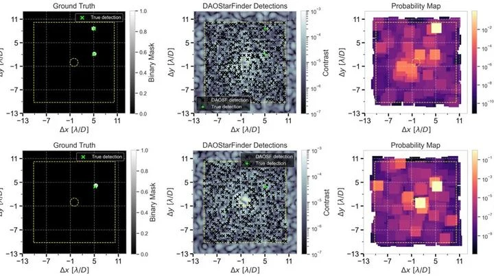
A pebble on a streambed lies still as sunlight sparkles dance on the water's surface. In astronomy, the contrast is similar: a real exoplanet is the stone, while myriad speckles (patches of scattered starlight) are the ripples trembling in instruments.
Genes are like houses in a city: in some, life bustles; in others, silence reigns. This 'urban' genetic landscape was analyzed in a thousand women with breast cancer.
A single-pixel camera works like an artist creating a picture through a stencil with many holes. In each frame, only a portion of the light, modulated by a mask, a spatial light modulator, reaches the single sensor, and then the computer reconstructs the full image by assembling this mosaic with computational algorithms.
Imagine a smart antenna panel, a phased array, that steers radio waves traveling at the speed of light. It works like an orchestra: hundreds of tiny emitters, like musical instruments, shift their phase (the moment they 'join' the overall sound) on command via a variable voltage.
Imagine a kinked garden hose: water meets resistance, and pressure drops. A similar process occurs in arteries when they lose their straight geometry. Vascular tortuosity is not just a cosmetic defect but a factor that disrupts hemodynamics and poses a threat of ischemic stroke.
Ultrasound waves bounce off organs to draw their outlines, but on an ovarian scan it's hard to tell a harmless fluid-filled cyst from a solid cancerous mass. Even specialists draw the boundaries differently, and this directly affects treatment choices.
The OvAi Focus program tackles this by learning from thousands of examples.
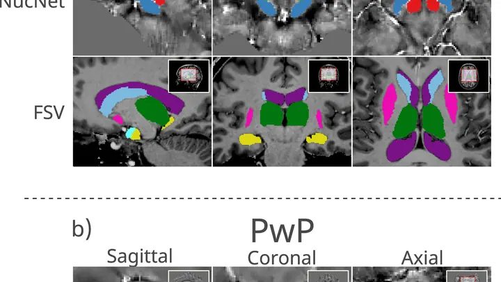
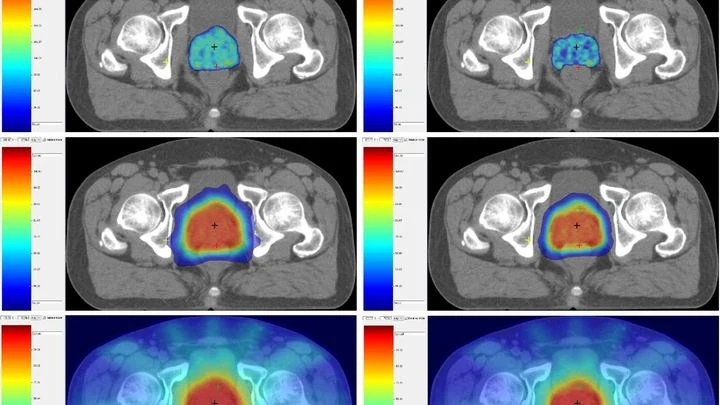
Prostate MRI scans often look like photos taken through a dirty window: noise, blurred edges, curvature from field imperfections. Scientists checked what happens if you 'wash' that window: remove the excess, correct the distortions, and accurately align different types of scans.
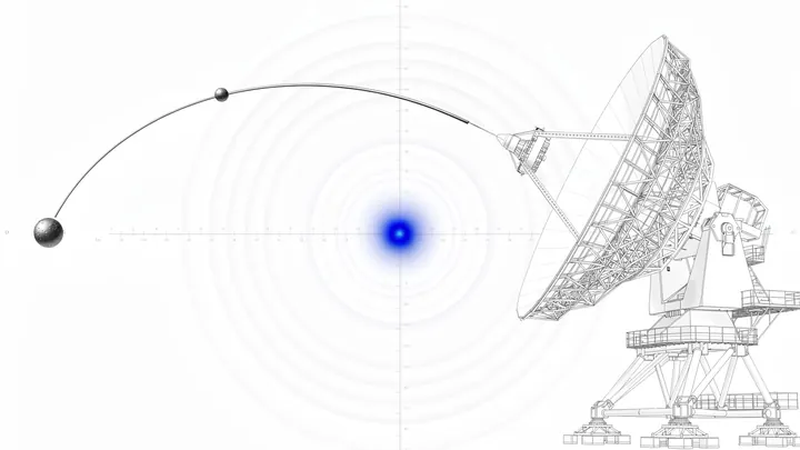
3I/ATLAS burst into the Solar System like an uninvited guest, a gift of fate for hunters of extraterrestrial intelligence. This comet-like wanderer from the depths of the Galaxy flew close enough to be heard in all its details — unlike distant exoplanets.
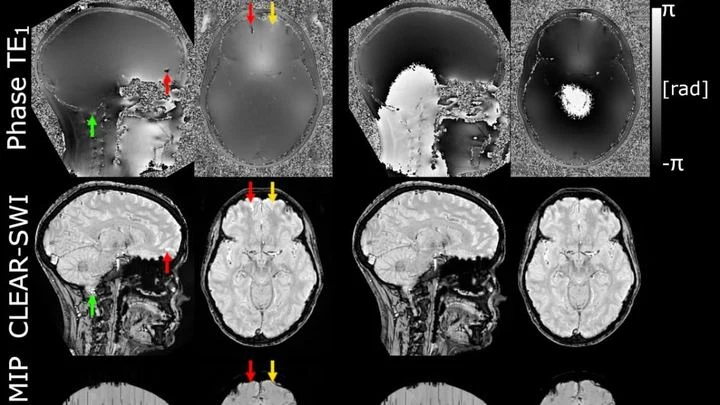
The powerful magnet of an MRI scanner induces weak electric currents in tissues. Their phase behaves like a compass needle trembling with every gust of wind — the slightest disturbance turns the map into a ripple, where instead of small vessels or iron deposits, all you see is noise.
For one person, the disease begins with a tremor in the hand — the cup dances on its saucer as they carry it to the table. For another, the legs suddenly refuse to move smoothly: the step becomes short and shuffling, as if the floor were suddenly covered in a sticky layer. These are not two different diseases but two faces of the same one — Parkinson's disease.
When you need to see inside the brain, you usually lie in a tight tube of an MRI scanner or get zapped with X-rays. Both methods are costly, bulky, and not always available.
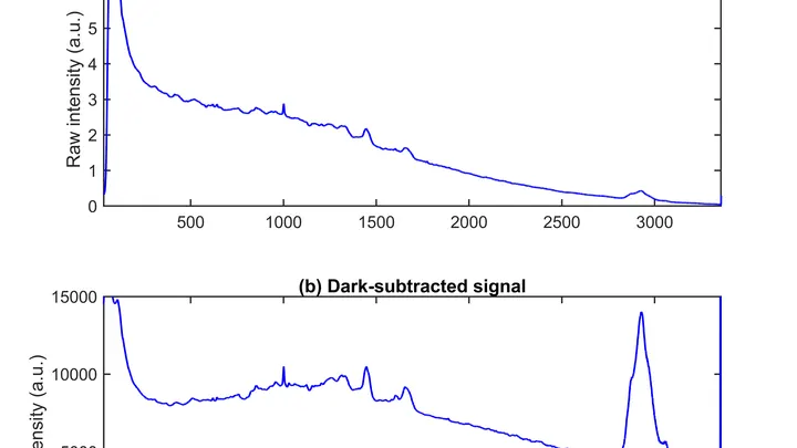
A drop of blood from a vein falls under three beams, and each one drags its own clues into the light. The infrared beam makes molecules vibrate — from that trembling, you learn how much sugar and fat is in the sample. A green laser scatters off proteins and lipids, shifting color — revealing the remodeling of cell membranes.
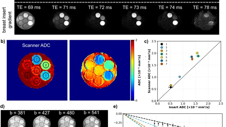
A flashlight spreads light in a broad beam, while a laser cuts a narrow path and picks out the finest details. In magnetic resonance imaging (MRI), the gradient, a rapid change in the magnetic field along the body, plays the role of that laser.
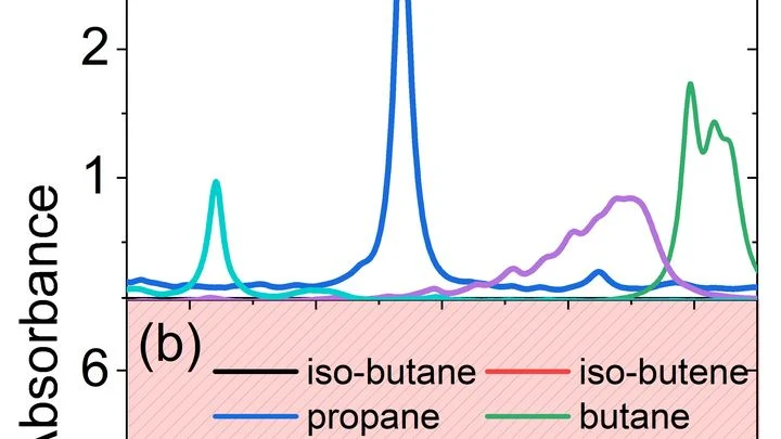
A hundred people are talking at once in a large hall. You try to hear one, but voices blend, some whisper, and you don't even suspect the existence of others. That's roughly how a conventional laser gas analyzer feels when dozens of substances mix in a combustion chamber or a factory pipe.
The light bulb turns on only in the room where you need it. That's roughly how photoactivatable drugs work: they 'switch on' only where light is directed, leaving the rest of the body untouched. This is especially important in cancer treatment: conventional chemotherapy hits all dividing cells indiscriminately, causing severe side effects.
An X-ray of a knee with osteoarthritis looks like a road map where the roads have narrowed and bumps have grown along the shoulders. Doctors look at this map and assign a grade from zero to four — how worn out the joint is. The problem is that two experienced specialists often see the map differently: one gives a three, the other a four.
Two specialists look at the same breast image. One is an experienced doctor, the other is an artificial intelligence program. Sometimes their opinions match, sometimes they diverge. How often they agree determines how accurately they can together distinguish healthy tissue from a tumor.
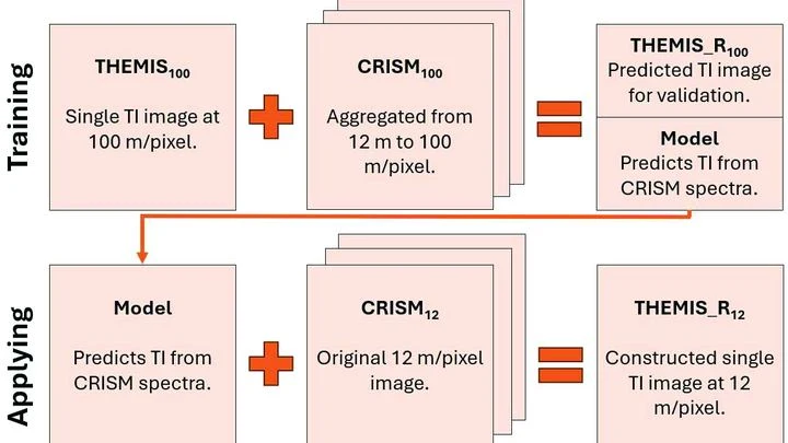
Warm rock and cold sand cool at different rates at night. An instrument orbiting Mars measures this heat, revealing whether the surface is loose dust, a dense crust, or solid rock. But thermal images have a resolution of 100 meters per point: an entire football stadium fits into a single pixel. Fine details (the beds of ancient streams, the edges of lava flows) are simply invisible.
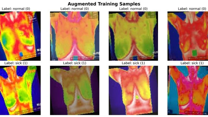
The hot air above asphalt shimmers, betraying the heated surface. Medical thermography works in a similar way: the skin over a tumor is often slightly warmer than healthy skin — by just fractions of a degree. Computer programs try to catch this difference and determine whether it's dangerous.
When a person's brain tumor is irradiated, some of the energy also hits healthy cells. A few months later, MRI scans of some patients show bright spots — places where tissue has changed due to radiation. Doctors call them "radiation-induced contrast enhancements." These spots aren't always dangerous, but they show where the brain took the hardest hit.
When a doctor irradiates a tumor, they try to spare nearby healthy organs. In the past, the standard approach was to give many small doses of radiation, like watering a lawn with a fine-spray watering can. Now, hypofractionation is increasingly used: fewer sessions, but larger doses, like a high-pressure hose. The question is how to tell whether the neighboring garden beds got overwatered.
At the Large Hadron Collider, protons collide, and from the flash dozens of particles are born. Among them may be traces of rare processes — for example, the decay of a Standard Model Higgs boson into two W bosons. But these particles fly apart so quickly that their tracks merge into a single wide jet.
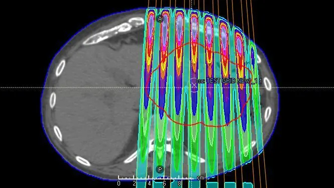
When treating large tumors, a conventional solid radiation beam can damage surrounding healthy tissue. Doctors came up with the idea of splitting the beam into many thin streams, like water from a watering can with fine holes. This approach is called spatially fractionated radiation therapy, or GRID therapy.
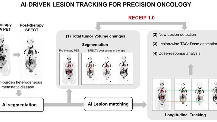
When treating prostate cancer, doctors often see not one tumor but many lesions in bones and lymph nodes. Each lesion can respond to therapy differently: one shrinks, another stays the same, a third grows. Previously, the disease was assessed as a whole, like judging the weather by the average temperature over a year.
In a hospital, blood from a finger prick usually goes to the lab, and the result comes back the next day. The new method promises to cut that wait to a few minutes. Instead of catching light reflected off a sample, it reads the electrical voltage that arises at the boundary between two media, like the faint tingle you feel when you touch a battery with wet fingers.
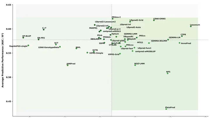
Geneticists read DNA like a long list of letters. From it, they compute a "polygenic risk score" — a number that shows how much higher or lower a person's chance of getting a disease, like diabetes, is compared to average.
PET/CT is like a map where cancer cells light up as bright spots. But what if the map is blank and the disease is still hiding somewhere? In 132 men with recurrent prostate cancer, doctors found no glowing lesions, but they decided to study the texture of the scan itself, like an experienced tracker noticing bent grass where an animal has passed.
There's a lot hidden underground: water, ore, sand, clay. To figure out what's there, you don't have to dig. Just run a weak alternating current through the ground and listen to how the rock 'responds' at different frequencies. This technique is called spectral induced polarization.
A laser hits the sample, and molecules inside start to vibrate. This vibration heats the material by tiny fractions of a degree. Previously, such heating was considered a nuisance, but now it's used to read chemical composition, like guessing who's coming down the hallway by the sound of footsteps.
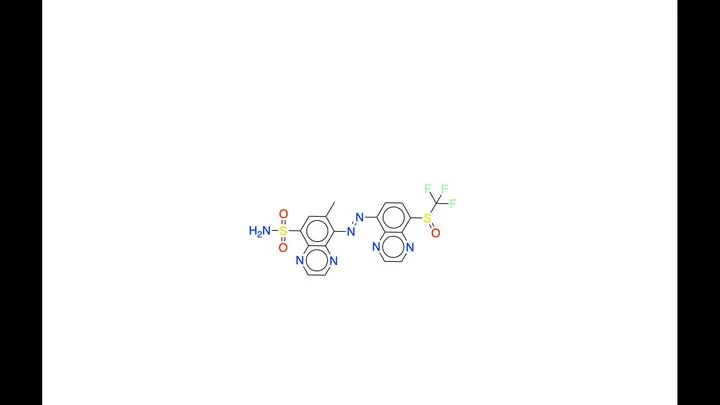
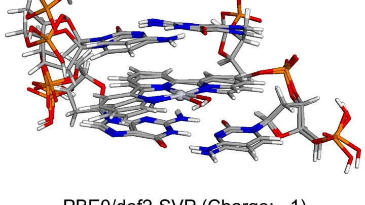
Standard chemotherapy hits the whole body like carpet bombing: both cancerous and healthy cells suffer. Scientists are hunting for more precise weapons — molecules that light up when they slip into a cancer cell's DNA, revealing its location. Such glowing probes could help doctors spot tumors early and even destroy them with pinpoint accuracy, leaving neighboring tissue untouched.
A mass spectrometer works like a molecular shredder: it breaks a substance into charged fragments and weighs each one. From this 'fingerprint,' chemists can tell what was in the sample. But if the molecule is new and its fragments don't match anything known, the instrument just shows a set of peaks, and what lies behind them is a mystery.
The authors of the paper proposed a workaround.
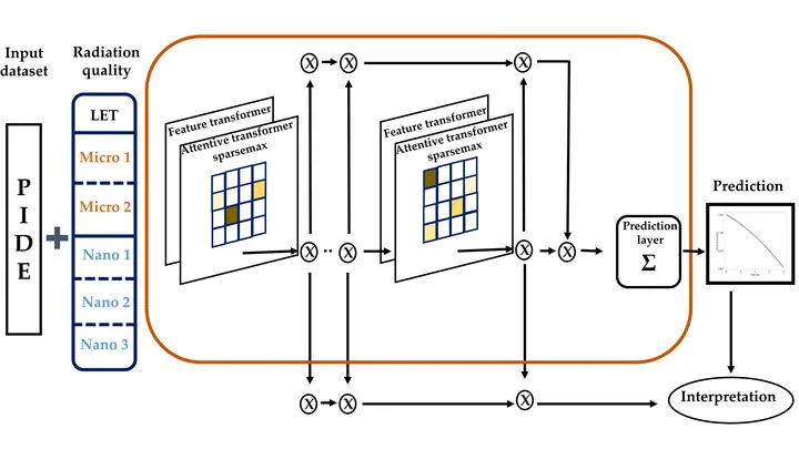
A cell hit by a proton beam is like a city after a pinpoint bombing: some blocks are leveled, others just have broken windows. Whether the city survives depends on which buildings are hit and how the strikes are distributed.
Glioblastoma, a brain tumor, is a tricky foe: in some patients, the drug temozolomide works, while in others it barely helps. The difference often lies in a small stretch of DNA — the MGMT gene. If this region is chemically switched off (methylated), the tumor is worse at repairing its cells, and the drug hits harder.
A glass of water and a glass of jelly look the same but behave differently. So it is with prostate tissue: on a standard slide under a microscope, you can see both the tumor glands and the supporting connective tissue around them. Previously, pathologists focused only on the glands, treating the support as mere background.
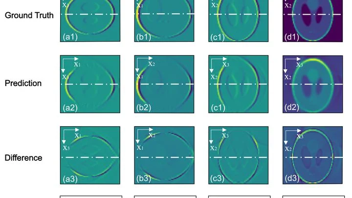
A laser hits tissue, and the tissue responds with a barely audible click — a sound wave. Sensors pick it up, and from that, an image of what's inside is built. That's how photoacoustic imaging works: light reaches deep, and sound carries the picture out.
In the new scheme, there are no sensors. Instead, several thin light beams are sent through the area where the wave has passed.
Many molecules, like amino acids, are like musical notes: their left and right versions sound almost identical, yet the difference is critical. One heals, the other is poison. Picking up this difference is hindered by inescapable noise, like in a room where a fan is loudly running. Regular spectroscopy misses the nuances.