Tumor imaging and characterization involves using medical imaging techniques — such as MRI, CT, PET, and optical spectroscopy — to non-invasively analyze the properties of tumors, including their shape, metabolism, cellular density, and microenvironment. The goal is to predict how a tumor will behave: whether it will grow aggressively, respond to chemotherapy, or resist radiation. This field bridges radiology, oncology, and computational biology, enabling personalized treatment planning based on each patient's unique tumor biology.
History
The field began with the clinical adoption of CT and MRI in the 1970s–1980s, but the term 'radiomics' was coined in 2012 by Philippe Lambin and colleagues, who proposed high-throughput extraction of quantitative imaging features. The integration of deep learning in the 2010s accelerated progress.
How it works
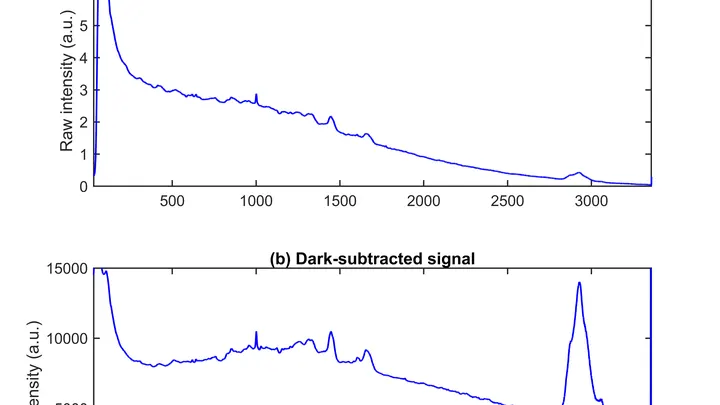
Different imaging modalities probe different aspects of tumor biology. MRI can map water diffusion (restricted in dense cellular regions) and perfusion (blood flow), while PET tracks metabolic activity using radiolabeled glucose. Advanced techniques like radiomics extract hundreds of quantitative features from images — texture, shape, intensity — and feed them into machine learning models that correlate with genetic signatures or treatment outcomes. For example, in glioblastoma, MRI-based features like necrotic volume and enhancing margin can predict response to chemotherapy before treatment begins. Optical spectroscopy and specialized microscopy (e.g., diamond-based sensors) can detect molecular changes at the cellular level, such as altered redox states or specific biomarkers.
In practice
Used in oncology clinics to guide biopsy targeting, predict treatment response (e.g., chemotherapy in glioblastoma), and monitor tumor recurrence non-invasively.
Fun fact
Tumor cells can migrate along white matter tracts in the brain, and advanced MRI techniques can visualize these 'invasion highways' to predict where a glioblastoma will spread next.
An old mattress stops bouncing back: the filling clumps up, and the springs lose their resilience. The ovary changes with age in much the same way, except that instead of filling, stiff connective tissue grows inside it, and the role of springs is played by the cells that support the eggs.
High-frequency sound focused on a single point acts like an invisible lens gathering sunlight. Inside a tumor, such a lens can raise the temperature to about 72°C in a second, like hot water from a tap, but inside living tissue. Meanwhile, surrounding healthy cells remain almost untouched.
An ordinary flashlight emits light whose waves oscillate in all directions at once. But pass that light through a special filter, and you get a beam that oscillates strictly in one plane: that's polarized light. When such a beam travels through biological tissue, the tissue's internal structure slightly alters the orientation of the oscillations.
A delicate watermark on wet paper, and a fogged-up glass before your eyes. That's roughly what reading quantum states of light looks like: fragile photon patterns get distorted by losses, and detectors catch only a small fraction of the signal.
Genes are like houses in a city: in some, life bustles; in others, silence reigns. This 'urban' genetic landscape was analyzed in a thousand women with breast cancer.
Imagine a kinked garden hose: water meets resistance, and pressure drops. A similar process occurs in arteries when they lose their straight geometry. Vascular tortuosity is not just a cosmetic defect but a factor that disrupts hemodynamics and poses a threat of ischemic stroke.
Medical scans are maps of tissue density. The brightness of each pixel reveals what’s there, much like photometry measures light intensity. But conventional algorithms analyze pixels in isolation, producing jagged organ boundaries.
A new approach compels the program to prioritize smoothness.
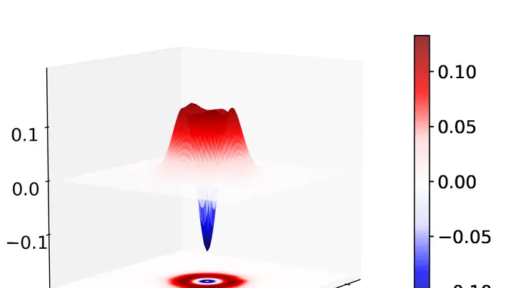
Each positron annihilation is like the birth of a pair of dancers, connected by an invisible thread of polarization. Two gamma quanta fly apart, but their states remain entangled: measuring one instantly determines the other — as if one partner senses every movement of the other, no matter the distance.
A radiologist stares at gray-white smudges on the screen — dozens of body slices where every suspicious spot must be found. Manual segmentation of one patient takes half an hour, and a large clinic sees hundreds of such patients. Fatigue and varying experience inevitably introduce variability: two specialists will outline the same lesion slightly differently.
When you need to see inside the brain, you usually lie in a tight tube of an MRI scanner or get zapped with X-rays. Both methods are costly, bulky, and not always available.
A drop of blood from a vein falls under three beams, and each one drags its own clues into the light. The infrared beam makes molecules vibrate — from that trembling, you learn how much sugar and fat is in the sample. A green laser scatters off proteins and lipids, shifting color — revealing the remodeling of cell membranes.
An X-ray tomograph is like a carpenter who examines a log from all sides to see where the void is inside. It takes dozens of flat shadow images, rotating the source and detector around the object. Then a 'reconstruction' program glues these shadows into a 3D model. The quickest reconstructor is the FBP algorithm (filtered back projection).
To peek inside a muscle, it is illuminated with radio waves in a strong magnetic field: that's how MRI works. Water in cells and water in blood respond differently, but a standard scanner doesn't tell them apart: it produces an averaged image where the signal from capillaries is superimposed on the signal from muscle fibers.
The light bulb turns on only in the room where you need it. That's roughly how photoactivatable drugs work: they 'switch on' only where light is directed, leaving the rest of the body untouched. This is especially important in cancer treatment: conventional chemotherapy hits all dividing cells indiscriminately, causing severe side effects.
A tongue tumor is like a stranger in a crowd. On the surface, they all look much alike, but beneath the surface hide very different personalities. Some tumors behave aggressively, others grow slowly. Doctors need to understand who they're dealing with before surgery.
Usually, this means taking a tissue sample — a biopsy.
A doctor studies a brain scan: somewhere in the tangle of tissue, a tumor is hiding. One glance picks out small spots and edges, another takes in the overall shape and location. A person combines both, but across hundreds of scans that gets tedious. A new program does the same thing automatically.
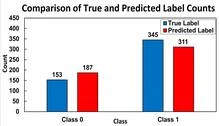
Two specialists look at the same breast image. One is an experienced doctor, the other is an artificial intelligence program. Sometimes their opinions match, sometimes they diverge. How often they agree determines how accurately they can together distinguish healthy tissue from a tumor.
A surgeon removes a breast tumor, but how do you know no cancer cells are left at the edge? Right now, the tissue goes to a lab, and the answer comes back days later. If the margin is not clean, a second surgery is needed. The new method promises to give the answer right in the operating room, while the patient is still on the table.
Inside every cell, there are tiny power stations — mitochondria. They burn nutrients and store energy. Two helper molecules, NADH and FAD, oversee this process. Previously, they were detected by their faint glow, but it was dim, and signals from different molecules overlapped.
The new microscope works differently.
A mole on your skin looks like a mosaic of colored pieces. Each piece is a small patch of the image where color and texture barely change. Such pieces are called superpixels. If you connect them with lines, you get a network that looks like a subway map: stations are the pieces, and lines are the connections between them. The weight of each line shows how strongly two pieces are related.
The radioactive isotope terbium-149 acts like a tiny homing bomb: it latches onto cancer cells and detonates with alpha particles that destroy the tumor from within, barely touching healthy tissue. But this bomb has a quirk: it also glows with positrons, and that glow can be captured by a special camera.
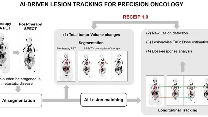
When treating prostate cancer, doctors often see not one tumor but many lesions in bones and lymph nodes. Each lesion can respond to therapy differently: one shrinks, another stays the same, a third grows. Previously, the disease was assessed as a whole, like judging the weather by the average temperature over a year.
An X-ray image is like a photo taken in the dark: the less light, the harder it is to see details. Doctors wanted to know how much you can dim the "light" in computed tomography (CT) and still not miss a disease. To do this, they took 50 pairs of chest images: one with a full radiation dose, the other with a low dose.
PET/CT is like a map where cancer cells light up as bright spots. But what if the map is blank and the disease is still hiding somewhere? In 132 men with recurrent prostate cancer, doctors found no glowing lesions, but they decided to study the texture of the scan itself, like an experienced tracker noticing bent grass where an animal has passed.
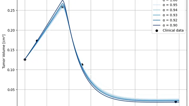
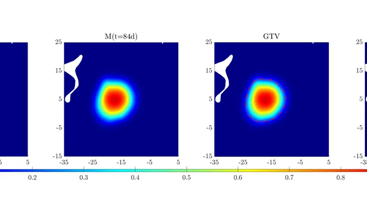
A tumor doesn't grow like a tree; it grows like a city: today's skyline is shaped by yesterday's construction and the fires of the day before. Standard tumor growth equations don't account for this: they only look at the current moment, as if the city were rebuilt from scratch every day. The authors added "memory" to the model: now the equation remembers what came before, and that changes the forecast.
A lightning bolt strikes an anthill. Some ants die instantly, some freeze, and a few days later the survivors start rebuilding the tunnels. That's roughly how a tumor behaves after electroporation — a method where short electric pulses are passed through tissue to poke holes in cancer cell membranes and kill them.
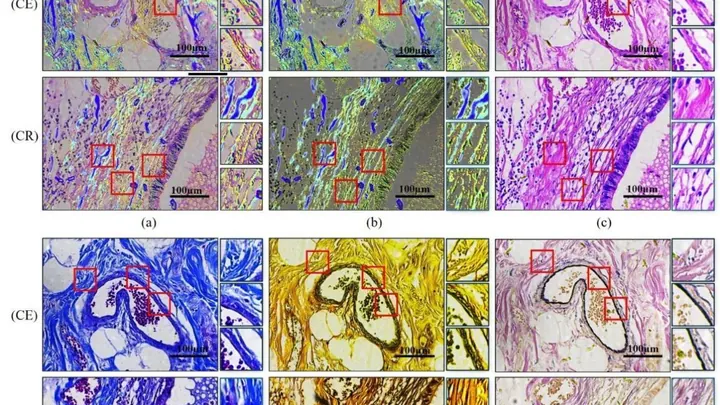
Inside our bodies, between cells, there is a support network — the extracellular matrix. It's like scaffolding made of proteins and carbohydrates that holds cells in place. When the fibers in this network change, it can be a sign of disease — for example, cancer or fibrosis.
Usually, to see these fibers, tissue is cut into thin slices and stained with special dyes.
A brain tumor is like a traveler trudging through a dense forest: trees and bushes are the different types of brain tissue, some easier to traverse than others. Glioma cells don't move in a straight line; they constantly loop around, bumping into obstacles and going around them.
In a heart-lung machine, blood flows through narrow tubing and past rapidly spinning pump blades. Sometimes red blood cells can't take it: their membranes rupture, and hemoglobin spills into the plasma. For a patient, this is like pouring sand into a car's fuel system — small damages accumulate and wreck the whole mechanism.
A tumor in the kidney grows like a weed that has learned to hide from the gardener. The immune system, that very gardener, simply doesn't notice it. Doctors try different drugs, but each person's disease behaves differently.
The authors made a computer replica of this battle.
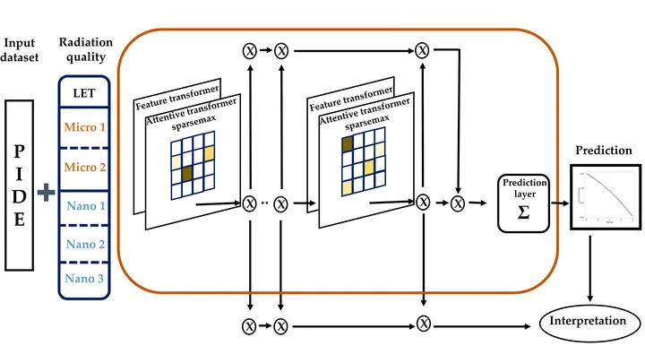
Treating cancer with a proton beam is a bit like archery: you need to hit the bull's-eye without hitting anything else. Protons release their energy at the end of their path, so healthy tissues are largely spared. But to make the shot safe, every treatment plan is checked in advance — measuring the dose the tumor will get and what the surrounding organs will receive.
A cell hit by a proton beam is like a city after a pinpoint bombing: some blocks are leveled, others just have broken windows. Whether the city survives depends on which buildings are hit and how the strikes are distributed.
A drop of blood can reveal how a tumor in the lungs is behaving. Doctors have long noticed that when the disease retreats, some blood markers rise while others fall. But tying these changes into a single coherent picture has been elusive.
Glioblastoma, a brain tumor, is a tricky foe: in some patients, the drug temozolomide works, while in others it barely helps. The difference often lies in a small stretch of DNA — the MGMT gene. If this region is chemically switched off (methylated), the tumor is worse at repairing its cells, and the drug hits harder.
A glass of water and a glass of jelly look the same but behave differently. So it is with prostate tissue: on a standard slide under a microscope, you can see both the tumor glands and the supporting connective tissue around them. Previously, pathologists focused only on the glands, treating the support as mere background.
Usually, to understand a quantum state of light, you need to perform dozens of complex measurements, like trying all the keys on a keychain. The new AI model works differently: it's a universal key that, after a slight adjustment, opens many 'locks'—simple and the most tricky states.
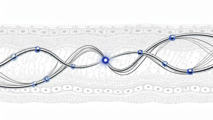
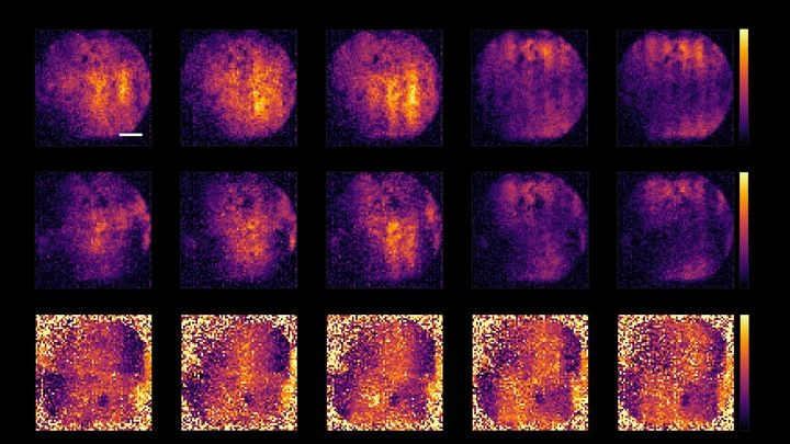
Every cell is a miniature tuning fork. A sharp magnetic pulse makes it 'ring', and the decay of that sound depends on what's inside: viscosity, density, composition. A tiny diamond with a structural defect acts like a hypersensitive microphone, picking up magnetic echoes from individual cells.
A brain tissue slice is just 5 microns thick, twenty times thinner than a human hair. On such a slice, a pathologist looks for signs of a tumor: altered nuclear shapes, cell crowding, abnormal blood vessels. It is painstaking work, and two specialists can disagree on the same image.