Carbon Beam Under Control: How to Catch a Gamma-Ray Flash express
When a carbon ion beam enters the body, it doesn't release energy all at once but rather at the end of its path, like a runner who gives their all on the final stretch. Doctors need to know this finish point to avoid damaging healthy tissue. But how can you look inside the body without making an incision? Physics itself provides the answer: when ions collide with atomic nuclei, brief flashes of gamma radiation are produced.
The idea is simple: if you record the moment an ion enters and the moment a gamma-ray flash arrives, the time difference tells you how deep the collision occurred. This is like an echo in the mountains — by the delay of the sound, you can tell how far away the cliff is. Scientists have tested this method for the first time on a carbon ion beam at the Italian CNAO center. They irradiated a plastic target 30 centimeters thick and caught the gamma rays with a detector placed to the side of the beam.
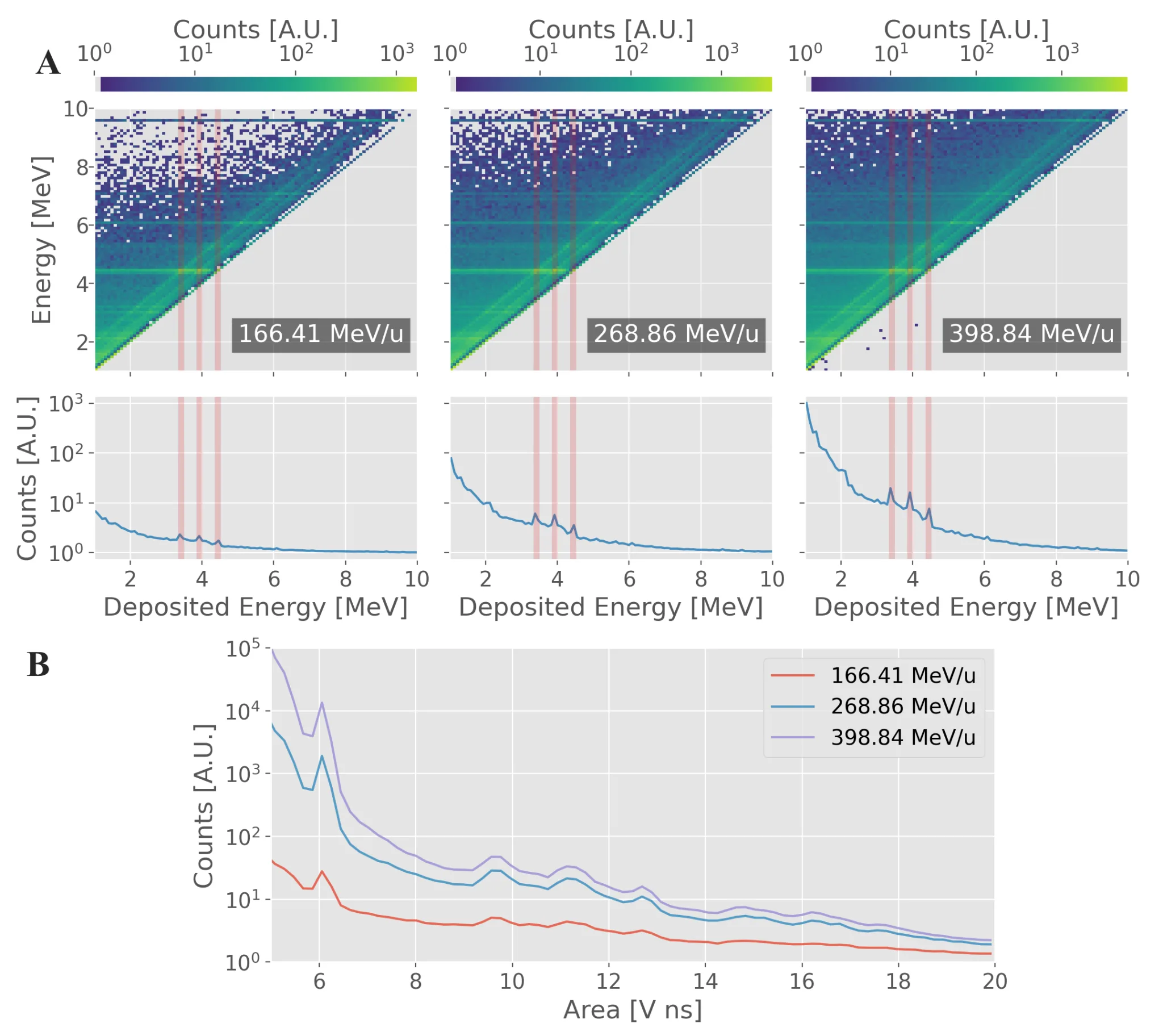
Experiments at three different beam energies showed that calculations and reality match almost perfectly. For the lowest energy, there was no discrepancy; for the middle energy, less than one percent; and for the highest energy, up to eight percent in the worst case. Moreover, the signal was clean: it was mostly gamma quanta, not interfering neutrons or nuclear fragments. This means the method can be trusted.
Why is this needed? Today, when treating with particle beams, doctors rely on calculations, but the patient's body is not perfectly uniform, and the beam may stop slightly earlier or later. If we can learn to track the depth in real time, treatment will become more precise and safer. For now, this is a laboratory experiment, but it proves that the echo from gamma-ray flashes could become a reliable compass for radiation therapy.
Original abstract
as written by the authors arXiv:2604.16131
Prompt Gamma Timing (PGT) is a promising technique for in vivo range verification in particle therapy, exploiting the time-of-flight between primary particles and prompt gamma rays emitted by nuclear interactions. PGT distribution is highly sensitive to beam energy and target density, which, under controlled detector positioning, enables real-time monitoring of particle range, detection of morphological changes, and support for adaptive treatment strategies. This study investigates for the first time the application of PGT in carbon ion therapy. Measurements were performed using a dedicated detection system composed of a silicon strip sensor for primary ion timing and a LaBr3(Ce) read out by a SiPM for secondary radiation. Carbon ion beams with energies of 166.41, 268.86, and 398.84 MeV/u irradiated a homogeneous 30.0 cm PMMA target at CNAO. The secondary radiation detector was positioned at four off-beam positions to assess the robustness of the PGT technique. Simulations based on Geant4 were conducted for all configurations to evaluate agreement and predictive capability. A bin-by-bin comparison of experimental and simulated PGT intensities demonstrated strong agreement within the 95% confidence interval, with no incompatible bins at 166.41 MeV/u, at most 1% at 268.86 MeV/u, and up to 8% at 398.84 MeV/u, depending on detector position. Photons were identified as the dominant contribution to the detected signals, particularly for detector positions upstream with respect to the primary particle beam, minimizing signal contamination from neutrons and charged fragments. The validated experimental-simulation framework confirms the capability of the proposed PGT system to resolve energy-dependent differences and highlights its potential for detecting clinically relevant changes in the particle beam range, supporting further development toward real-time monitoring in carbon ion therapy.
Gamma-ray flashes from carbon irradiation occur within billionths of a second after the ion hits — faster than the blink of an eye.