How a Cell Makes a Waist express
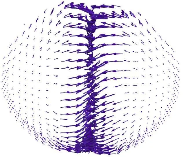
A cell about to divide first marks its waist. Beneath its membrane, in a thin gel of protein filaments, movement begins. The filaments flow like a slow river, and a belt forms in the middle of the cell. This belt tightens more and more until the cell is pinched in half.
Scientists built a computer model of this process. They saw that the ring of filaments forms on its own if the filaments are initially aligned in one direction. Even without an external command. But if the filaments are arranged randomly, no ring forms. So the cell remembers how its filaments were arranged before division.
This result helps explain why cell division sometimes goes wrong. If the filaments are not arranged properly, the ring won't form, and the cell won't be able to divide. And division errors underlie many diseases, including cancer.
Original abstract
as written by the authors arXiv:2604.17349
During cell division active flows occur in the cortex, a thin layer of gel like network of acto myosin filaments, beneath the cell surface. The cortical flow and the associated stresses bring about change in the cell shape, in particular a sharp invagination at the mid cell. Using 3D phase field simulation of an active deformable shell, which captures coupled dynamics of cortical velocity and nematic order, we show how a nematic like actomyosin ring spontaneously emerge at the equator and drive sharp invagination. We further demonstrate how different cortical flow patterns, including counter rotating flows emerge near the division furrow. We show that these flow patterns, often attributed to intrinsic chirality of actomyosin filaments can instead arise from bias in the initial nematic alignment, revealing a memory effect in the system. By analyzing a simpler model of activity gradient driven compressive flow on a flat interface we decipher the main ingredients for surface instability leading to invagination and counter moving flows.
The belt that divides a cell is less than a thousandth of a millimeter thick. But it can squeeze the cell with enough force to tear it into two parts.