X-ray microscope becomes three times sharper express
An X-ray beam can peer inside an opaque piece of metal and show where cracks or stresses are hiding. But to see such defects, the beam must be focused into a very narrow pencil, just as sunlight is focused by a magnifying glass into a hot spot. In an X-ray microscope, the lens plays the role of the magnifying glass. Only glass won't work for X-rays: the rays pass straight through it, barely bending. So scientists make lenses from many alternating layers of two materials — molybdenum and silicon. Each layer deflects the beam by a tiny angle, and together they focus it to a point.
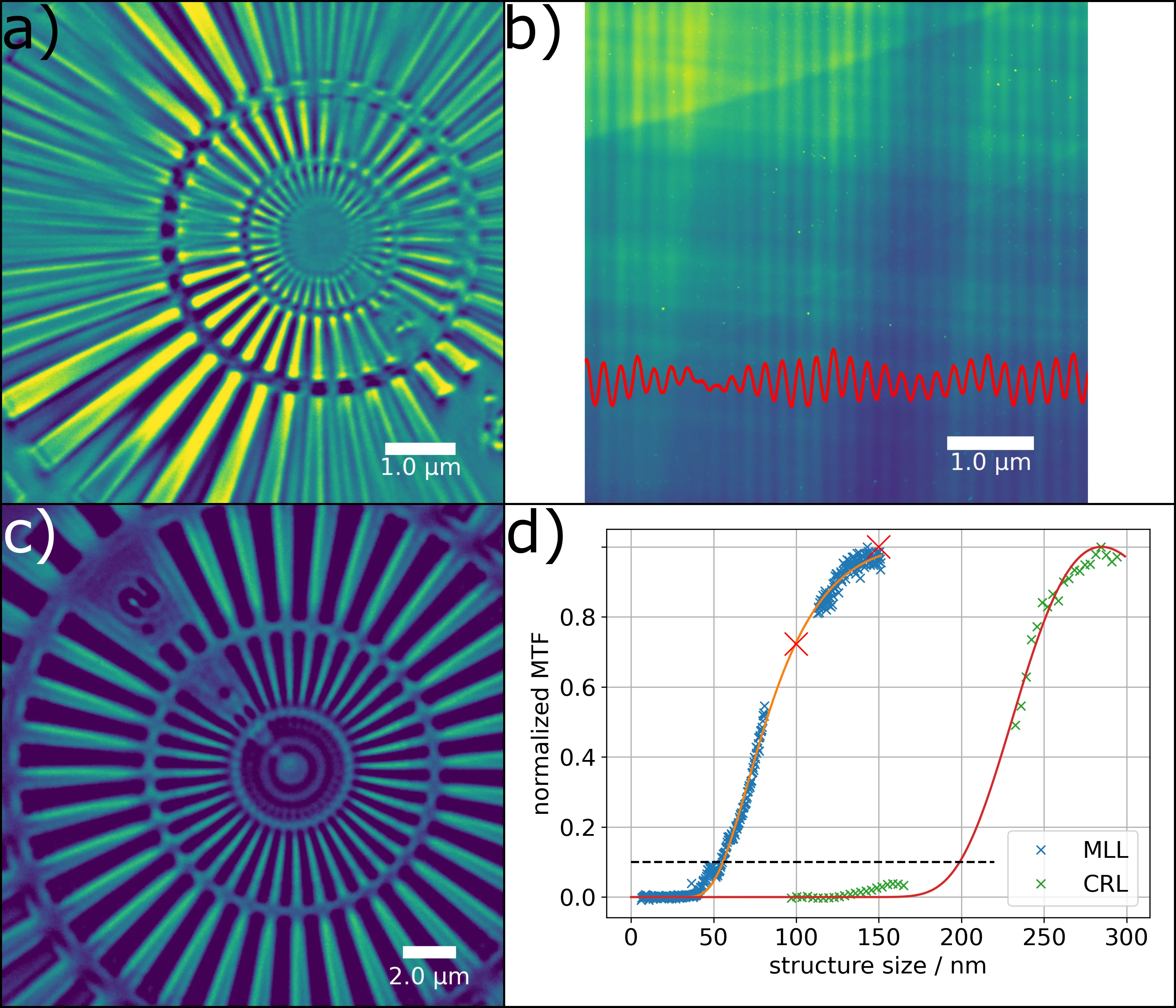
The authors of the new study placed not one such layered lens but two, crossed at right angles, in the X-ray microscope at the ESRF synchrotron in Grenoble, France. Each lens focuses the beam in one direction, and together they focus it into a small spot. The resulting spot was 56 nanometers across — three times smaller than what the previous plastic lens achieved. For comparison: the flu virus is about 100 nanometers in diameter, so the new microscope can resolve details half the size of a virus.
The new lens transmits 26.7% of the X-ray light — enough to produce bright, high-contrast images. The scientists tested the microscope on a silicon pillar that connects layers of a microchip (such pillars are called TSVs). They could see how the silicon atoms are arranged inside the pillar and spot places where the crystal lattice is slightly tilted. These tilts often herald cracks that could cause the chip to fail.
The new lens is made of alternating layers of molybdenum and silicon, each only a few nanometers thick, like a stack of thousands of sheets of paper, but each sheet is a thousand times thinner than a human hair.