X-ray microscope sees twice as clearly express
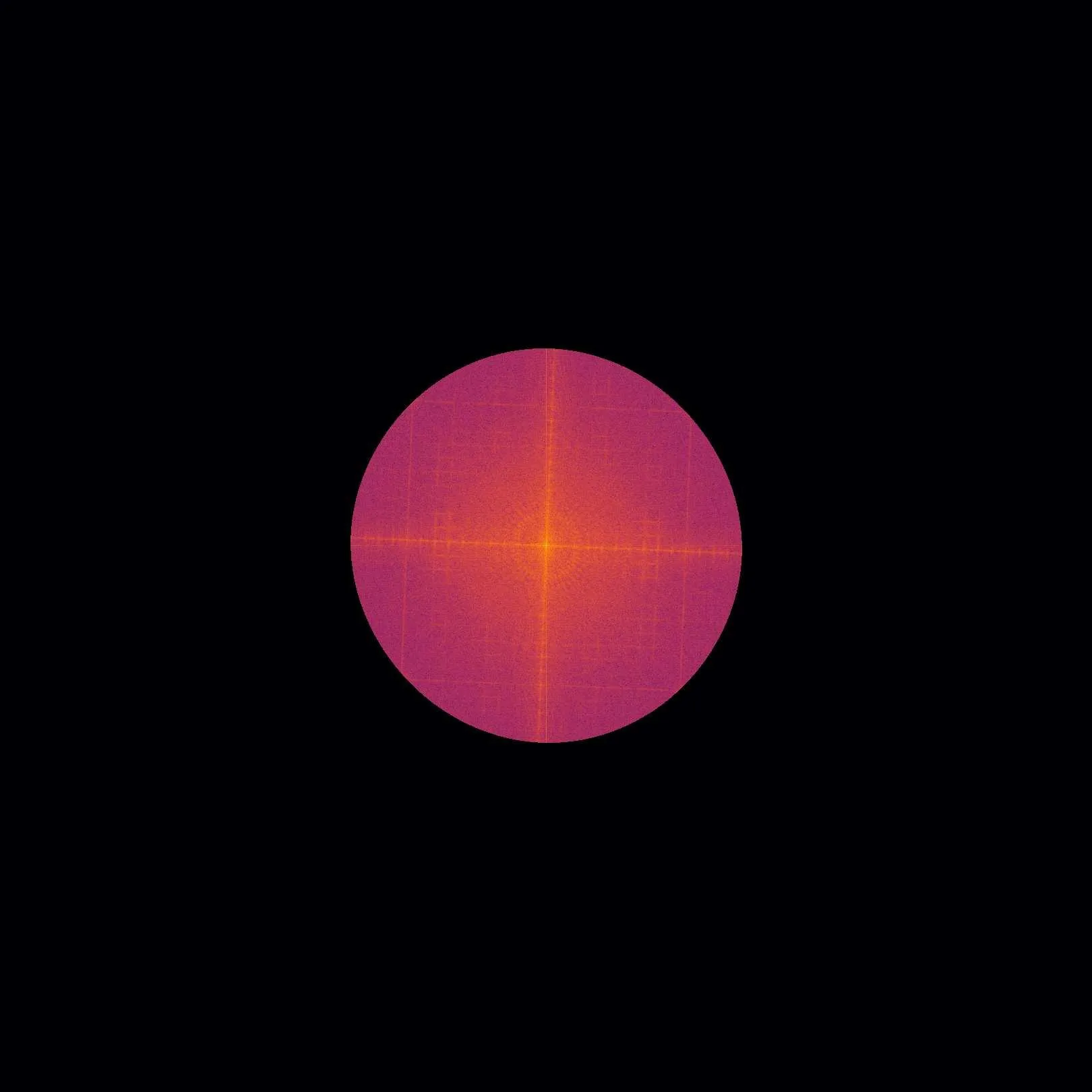
An X-ray image of a battery looks like a photo taken through frosted glass: fine details blur into a gray smudge. The culprit is the detector itself, which cannot distinguish features smaller than its pixel size. Engineers proposed getting around this barrier by illuminating the sample not with a uniform beam but with a striped pattern.
The stripes act like a stencil: they transfer information about the tiniest structures onto parts of the image that the detector sees well. By shifting the pattern several times and taking images, the computer decodes this data and assembles a picture with details twice as small as the previous limit.
The same principle applies not only to standard X-rays but also to phase contrast and dark-field imaging, which reveal details invisible to conventional methods. This paves the way for materials testing and medical imaging with details that were previously out of reach without bulky setups.
A 2.2-fold improvement in resolution means that on the same detector you can now see details that previously merged into a single pixel.