Heart by Contour: How AI Builds 3D Shape express
The heart is not a perfect sphere; its shape changes with every beat. To build an accurate 3D copy for a computer model, doctors typically take dozens of MRI slices. But that's like assembling a watermelon from thin slices: the fewer slices, the more guesswork.
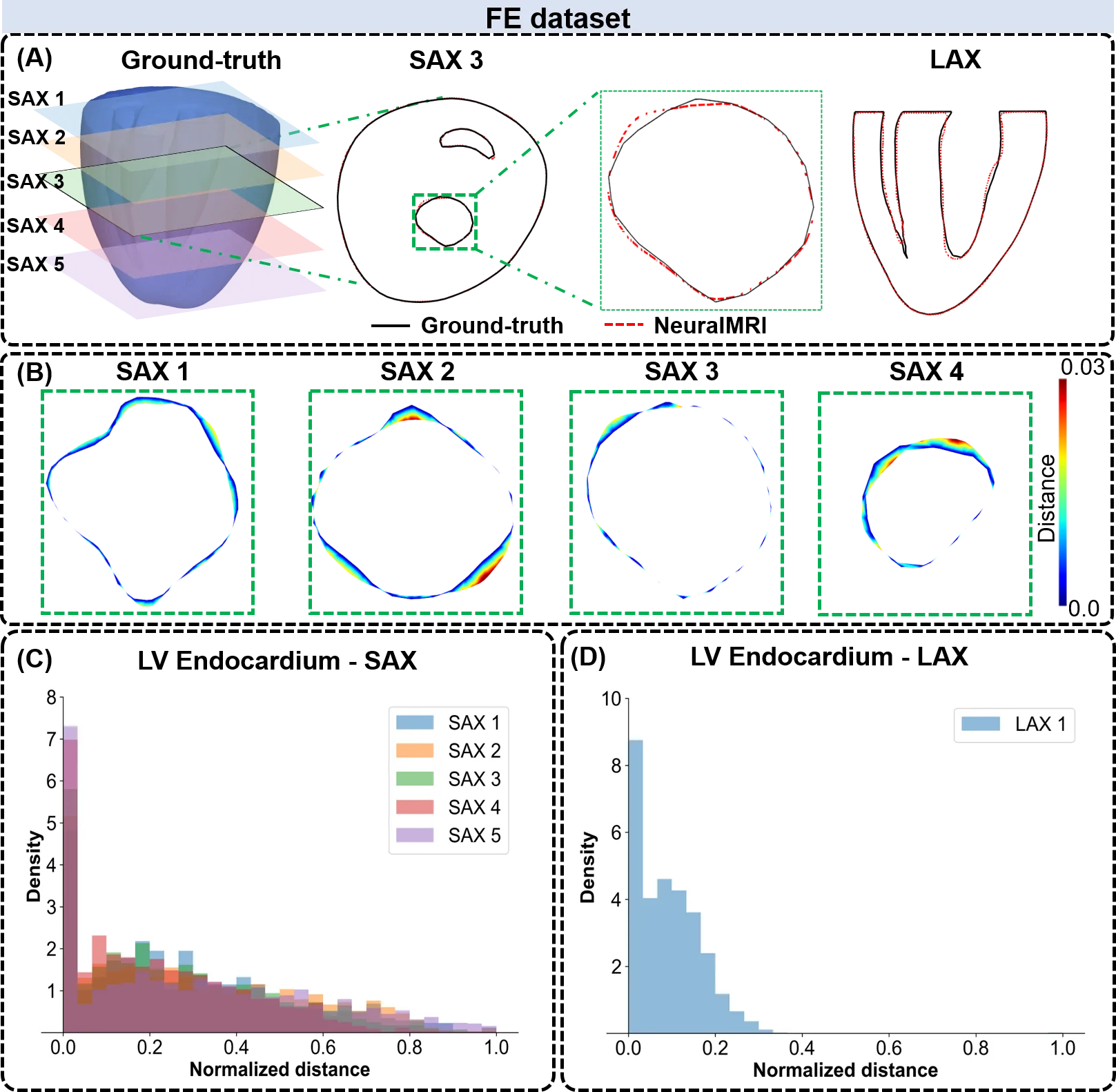
A new approach teaches a neural network to understand the heart's shape from sparse contours. It's similar to how a sculptor reconstructs a statue from a few sketches from different angles. Instead of rigid interpolation rules (drawing lines between points), the network learns what healthy and diseased hearts look like and 'fills in' the missing parts. In tests on computer models and real scans, the method reproduced the shape more accurately, especially at the heart's apex — a spot where conventional algorithms often stumble.
Such reconstruction is a first step toward a digital heart twin: a virtual copy on which drugs or surgeries can be tested before the real procedure. Scientists already use similar approaches for MRI of small animals, where every scan is precious.
Original abstract
as written by the authors arXiv:2603.27003
Computational models of cardiac structure and function are increasingly central to the development of subject-specific cardiac digital twins, enabling improved characterization of contractile dysfunction, pathological remodeling, and electrical abnormalities. A critical prerequisite for these models is the accurate reconstruction of three-dimensional (3D) cardiac anatomy from medical imaging. Multi-planar magnetic resonance imaging, particularly when combined with artificial intelligence, offers a clinically feasible alternative to conventional reconstruction techniques. In this study, we present a neural field-based reconstruction framework that recovers 3D cardiac geometries from sparse planar contour data by learning continuous shape representations. Reconstruction performance was evaluated using complementary in-silico and in vivo datasets spanning variations in sampling density and geometric complexity. Across both datasets, reconstructed meshes closely matched reference geometries, demonstrating that the neural field approach faithfully captures cardiac planar contours. Compared with traditional local interpolation methods, the proposed framework exhibited improved geometric fidelity in anatomically challenging regions, including the left ventricular apex and basal segments, particularly under sparse sampling conditions. Collectively, these findings demonstrate that neural field-based reconstruction provides a robust and efficient pathway for multi-planar cardiac shape recovery, with particular relevance for AI-driven modeling pipelines and data-limited settings such as small-animal and time-resolved cardiac imaging.
The apex of the left ventricle is one of the hardest spots for 3D reconstruction: it's pointed and moves a lot with each contraction.