Heart by Contour: How AI Builds 3D Shape express
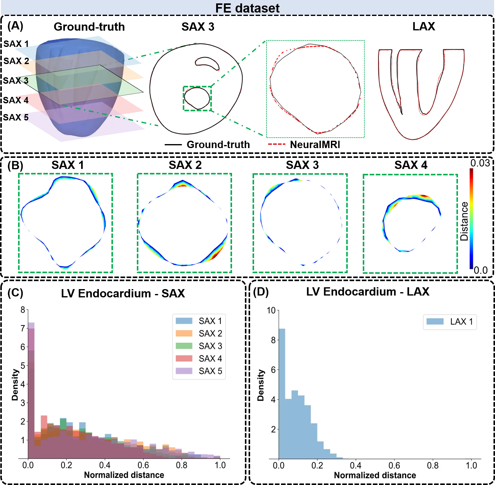
The heart is not a perfect sphere; its shape changes with every beat. To build an accurate 3D copy for a computer model, doctors typically take dozens of MRI slices. But that's like assembling a watermelon from thin slices: the fewer slices, the more guesswork.
A new approach teaches a neural network to understand the heart's shape from sparse contours. It's similar to how a sculptor reconstructs a statue from a few sketches from different angles. Instead of rigid interpolation rules (drawing lines between points), the network learns what healthy and diseased hearts look like and 'fills in' the missing parts. In tests on computer models and real scans, the method reproduced the shape more accurately, especially at the heart's apex — a spot where conventional algorithms often stumble.
Such reconstruction is a first step toward a digital heart twin: a virtual copy on which drugs or surgeries can be tested before the real procedure. Scientists already use similar approaches for MRI of small animals, where every scan is precious.
The apex of the left ventricle is one of the hardest spots for 3D reconstruction: it's pointed and moves a lot with each contraction.