X-Ray Vision Through a Sample Without Point-by-Point Scanning express
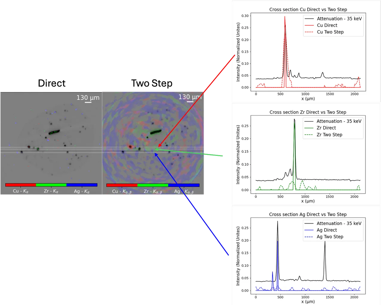
Conventional X-ray fluorescence analysis works like a meticulous scribe: to learn the composition of a picture, it walks over every square centimeter and logs whether lead or copper is present. For a flat sheet, that's tolerable, but for a three-dimensional object, it's torture. Tomography requires repeating the walk at many angles, and the number of measurements snowballs.
X-ray fluorescence occurs when an atom, excited by an X-ray beam, emits a characteristic glow with an energy unique to each element. From this glow, you can tell which elements are present and where. But if you measure point by point, a cube with a side of 140 voxels would require about 2.8 million individual measurements per angle.
The authors took a different approach: instead of a thin beam, they illuminate the sample with a broad pattern, like a random mosaic of bright and dark spots. The detector captures the total glow from the entire volume at once. By varying the patterns, they obtain a set of numbers in which information about all elements and all depths is mixed. Then a single mathematical algorithm untangles this knot and produces a three-dimensional map of element distribution.
This approach is especially valuable for large and heterogeneous samples — for instance, chunks of ore, biological tissues, or ancient artifacts that cannot be kept under the beam for long. The method requires no reconfiguration of the setup: the same sources and detectors as in conventional fluorescence tomography, just controlled differently.
The X-ray scribe has turned into a cryptographer: instead of visiting every point, it sends a series of patterns and reconstructs the entire book of composition from the response. For large tomographies, this cuts time from hours to minutes and opens the way to analyzing samples that previously simply couldn't be measured in time.
X-ray fluorescence can distinguish a fake from an original: old paints contain lead, mercury, or arsenic, while modern ones contain titanium and zinc. From the set of elements, art historians determine when and where a painting was made.
In the series 'Firefly,' a handheld scanner based on X-ray fluorescence instantly determines the composition of an alloy or mineral — the method in this article brings such imaging for volumetric objects closer to real-time.